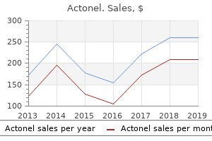
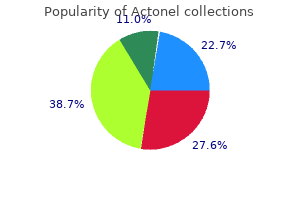
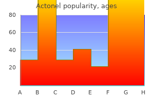
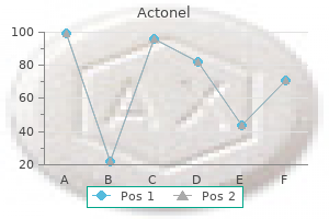
Actonel
Hitesh Kapadia, D.D.S. - Craniofacial Center, Department of Dentistry
- Seattle Children’s Hospital
- Seattle, Washington
Order 35 mg actonel overnight deliveryDegenerations most frequently begin in the peripheral cornea symptoms by dpo generic 35mg actonel otc, though central imaginative and prescient ultimately may be affected symptoms yeast infection women generic 35mg actonel mastercard. Inflammation sometimes is concerned early in the degenerative process and could additionally be accompanied by corneal vascularization medicine cups cheap 35 mg actonel visa. In some cases medications quizzes for nurses discount 35mg actonel with visa, these inflammatory processes are associated with systemic disease. The microcornea is generally clear, with normal histologic structure, and in the absence of other ocular abnormalities, imaginative and prescient could also be good. Numerous somatic abnormalities have been described along side microcornea and anterior microphthalmos, including dwarfism and Ehlers�Danlos syndrome. The diameter of the cornea is 13 mm or higher, however the corneal thickness and histologic anatomy are normal. Although X-linked recessive inheritance is commonest with 90% of all instances found among males, all modes of inheritance have been reported. Moreover, the megalocornea demonstrates regular endothelial cell population densities on specular microscopy, whereas in congenital glaucoma, these are diminished, ostensibly due to corneal distention. Both conditions, however, have been reported in the identical family and in the identical individual. True cryptophthalmos, also referred to as ablepharon, happens when the lids fail to kind. The cornea and conjunctiva are uncovered and endure metaplastic modifications to form pores and skin. The measurement of a normal new child cornea measures ~10 mm in horizontal diameter, whereas the scale of a standard adult cornea measures ~12 mm in diameter. The vertical diameter almost all the time is exceeded by ~1 mm by the horizontal diameter. The eye may be otherwise regular, but usually different ocular abnormalities, corresponding to colobomas, are current. Just as megalocornea is related often with anterior megalophthalmos, microcornea usually accompanies anterior microphthalmos, with crowding of the anterior segment structures generally leading to angle-closure glaucoma. The iris could exhibit transillumination defects because of attenuation of the dilator muscle. Because of the abnormal spatial relations of structures within the anterior segment and stretching of the zonules, iridodonesis, phakodonesis, and lens subluxation or dislocation may happen; the latter might end in secondary lens-induced glaucoma. Note the anterior phase with no abnormalities (except beveled scar of cataract incision and surgical aphakia). A number of pathogenetic theories have been advanced, all based mostly on concepts of anterior segment embryogenesis. The considerably archaic time period anterior phase cleavage syndrome, for instance, implies irregular separation of growing tissues. Arrest at any of these stages might bring in regards to the acknowledged medical dysgenesis syndromes. In addition to this developmental arrest, secondary anterior displacement of the lens-iris diaphragm may account for different abnormalities. The radius of curvature could reach ranges as low as 20�30 D, much like that of the sclera. In cornea plana, the anterior chamber is shallow by virtue of the low corneal dome. Refractive abnormalities vary from hyperopia of 7 D to myopia of 9 D, depending on the globe dimensions and corneal curvature. The distortion of the cornea, along with concomitant sclerocornea, leads to a decrease in corneal transparency. Most circumstances are sporadic, with both dominant and recessive inheritance pedigrees reported. The formation of the limbal anlage occurs between the seventh and tenth gestational weeks, permitting neural crest mesenchymal cells to differentiate into either sclera or cornea and to induce a corneal curvature that exceeds the scleral. Histopathologic research of sclerocornea have revealed morphologic options resembling scleral tissue. The stroma consists of irregularly organized collagen fibrils with an elevated diameter anteriorly, in distinction to the normal cornea. Gonioscopy shows that it juts into the anterior chamber, and the adjacent uveal trabecular meshwork may seem dense. Composite illustration of the anatomic findings in mesenchymal dysgenesis of the ocular segment. The markers in the table point out the corresponding anatomic element within the illustration. The central abnormalities happen due to focal absence or attenuation of the endothelium. The pneumotonometer or Tonopen is preferable to other applanation devices as a result of the presence of related corneal abnormalities or small radius of corneal curvature could give false intraocular stress readings. Assessment of the optic nerve is critical to determining the overall visual prognosis and deciding on the course of future therapy. Medical therapy could be helpful when intraocular stress is particularly high and temporizing measures are needed. This disorder has a usually poor surgical prognosis, each for glaucoma control and for corneal opacities, if current. Achieving a stability between persistent medicines and performing surgical procedure is uniquely tough. The introduction of effective use of antimetabolites for filtration in kids could favor of surgery when the optic nerve is threatened considerably. Nevertheless, this sort of remedy in kids stays a substantial concern because the eyes mature. Some attribute the cause to an irregular migration or differentiation of the secondary mesenchyme that normally forms the corneal stroma. In some cases, pigment surrounds the perimeters of the posterior despair, suggesting earlier contact to the iris. Eighty p.c of Posterior Keratoconus 500 Posterior keratoconus53�56 has no relation to anterior keratoconus. Left, Multiple facial anomalies similar to telecanthus, low nasal bridge, and maxillary hypoplasia. Right, this similar affected person reveals posterior embryotoxon, hypoplasia of the anterior iris stroma, corectopia, and peripheral anterior synechiae. Associated anomalies, corresponding to microcornea, sclerocornea, and infantile glaucoma, could additionally be present, however for the most part, no different ocular or systemic abnormalities are current. Centrally, the posterior cornea and lens could additionally be adherent, and there may be an anterior polar cataract. This kind extra generally is bilateral, and almost every concerned case reveals extreme ocular and systemic malformations. Other associated ocular abnormalities embrace microcornea, microphthalmos, cornea plana, sclerocornea, colobomas, aniridia, dysgenesis of the angle and iris and chronic hyperplastic major vitreous.
Diseases - Xanthic urolithiasis
- Gonadal dysgenesis, XX type
- Phenobarbital antenatal infection
- Sulfite and xanthine oxydase deficiency
- Dementia with Lewy bodies
- Myoclonus epilepsy partial seizure
- Alcohol fetopathy
Actonel: 35 mg
Purchase actonel 35mg overnight deliveryBacillus species are recognized in soil and have been linked to ocular infection after trauma due to symptoms 5 days past ovulation generic 35 mg actonel mastercard contaminated metallic overseas our bodies treatment 0f ovarian cyst discount 35 mg actonel overnight delivery. Its virulence could also be associated with the manufacturing of toxins together with phospholipases 4 medications at target order actonel 35mg with visa, proteases treatment innovations discount actonel 35mg with mastercard, hemolysins, enterotoxin, and emetic toxin. The presenting signs of bacterial keratitis differ depending on the virulence of the organism, period of infection, structural standing of the cornea, and host inflammatory response. Common presenting symptoms include ache, decreased vision, tearing, and photophobia. Eyelid edema, conjunctival hyperemia with a papillary reaction, and chemosis are typical findings. A corneal epithelial defect with adherent mucopurulent exudate and underlying stromal infiltrate is a trademark signal for infectious keratitis. Multiple focal infiltrates could be seen with contact lens use or with polymicrobial infections. Migration of inflammatory cells causes a diffuse mobile infiltration adjoining to the ulcerated stroma. An anterior chamber response can vary from gentle aqueous cells and flare to a marked hypopyon. A cornea broken from prior illness can current with much less distinct signs and signs. Preexisting corneal scars, epitheliopathy, or inflammation confuse the picture as do prior use of antibiotics and corticosteroids. On examination, all ocular abnormalities should be documented in detail to help track the medical course on subsequent visits. Trauma with vegetative matter, dirt, or gravel in addition to contact lens use are common predisposing factors. The clinical image resembles mycotic keratitis which may delay proper prognosis and remedy. Bacterial keratitis from Mycobacterium is caused by nontuberculous species, whereas major corneal an infection from Mycobacterium tuberculosis and Mycobacterium leprae is uncommon. Nontuberculous mycobacteria are cardio acid-fast rods that are ubiquitous and may be present in water, mud, soil, animals, milk, and different foodstuffs. These organisms have lipid-rich cell walls which contribute to their acid-fast staining characteristics. Nontuberculous mycobacteria cause an indolent keratitis normally occurring after trauma or surgery, including cataract extraction and penetrating keratoplasty, and barely with contact lens use. Unusual clinical presentations embrace a linear pseudodendritiform look, ring infiltrate, and crystalline keratopathy. The infiltrate, which may be multiple, begins in the interface and spreads to adjacent stroma of the flap and stromal mattress. In addition to a focal infiltrate, a cracked windshield appearance of infectious crystalline keratopathy has been reported. The flap ought to be lifted to get hold of sample until the infiltrate is in the periphery or has perforated through the flap. In addition to Gram and Giemsa stains, Ziehl�Neelsen stain to examine for acid fastness and fluorochrome stain which reveals yellow-orange fluorescence must be obtained to determine Mycobacterium on smears. Careful microscope diagnosis is warranted since misdiagnoses with Nocardia and Corynebacterium have occurred. Fast-growing Mycobacteria are tradition positive within 7 days whereas gradual growers require a quantity of weeks. Fortified antibiotics are made by mixing the powdered drug or diluting the parenteral form with synthetic tears or balanced salt answer. These freshly prepared options remain stable for up to per week without important lack of exercise. Although ointments prolong corneal contact time and lubricate the ocular floor, peak corneal concentrations could also be restricted when compared with solutions. Subconjunctival injections might not have a therapeutic advantage over topical options. Soft contact lenses and collagen shields can act as drug supply devices and help in sustaining high corneal drug levels. Systemic remedy is indicated for gonococcal infections as nicely as for younger children with severe H. Empiric Therapy Since bacterial keratitis can rapidly progress and threaten vision, therapy should be begun when an infectious course of is suspected. Topical broad spectrum antibiotics are initially used and later modified according to culture results, antibiotic susceptibilities, and scientific response. For severe cases, mixture remedy with fortified beta-lactam (cefazolin 50 mg/mL) and aminoglycoside (tobramycin or gentamicin 14 mg/mL) offers sufficient coverage of each Gram optimistic and adverse organisms that cause bacterial keratitis. Vancomycin (50 mg/mL) can be substituted for cefazolin in instances of penicillin allergy or resistance to Enterococcus and Staphylococcus species. Single-agent remedy with fluoroquinolones has been shown to be as efficient as combination remedy in treating bacterial keratitis. They require two mutations to establish resistance and, subsequently, are simpler towards Gram-positive organisms that already have a single mutation and are proof against older-generation fluoroquinolones. Positive indicators of medical improvement embrace decreased ache, decreased discharge, consolidation of the stromal infiltrate, decreased anterior chamber response, and corneal reepithelialization. Culture and antibiotic susceptibility outcomes can be utilized to focus remedy towards the offending organism or to discontinue unnecessary drugs. Clinical improvement will not be seen through the first 2 days because of increased irritation and suppuration from bacterial exotoxins. A lack of improvement or scientific worsening after 48 h could warrant repeat cultures, although concomitant antibiotic therapy will lower yields. Increased drug Bacterial, Chlamydial, and Mycobacterial Infections soaking of the stromal bed and flap with antibiotics. Maximal topical antibiotic remedy in addition to systemic antibiotics is given preoperatively. Corticosteroids may play a restricted position in treating bacterial keratitis with its potential for lowering the host inflammatory response and resultant corneal scarring. Adverse results of corticosteroids include inhibition of corneal wound healing, promotion of stromal thinning and perforation, potentiation of microbial replication and recrudescence of infection, secondary glaucoma, and cataract formation. Worsening or recrudescence of Pseudomonas keratitis has been reported after the addition of topical steroids. Pain management with analgesics may provide not solely consolation, but additionally increased compliance with the troublesome routine of across the clock topical drops. Cycloplegic brokers can be used to lower discomfort from ciliary spasm and to forestall synechiae formation. Cyanoacrylate glue can be used to reinforce an area of corneal thinning, a descemetocele, or a small perforation. This procedure permits for further remedy of the infection and irritation whereas postponing surgical procedure. Alexandrakis G, et al: Corneal biopsy within the management of progressive microbial keratitis.
Discount actonel 35 mg otcKeratometry measures often four factors on the central cornea 3 mm apart and assumes an everyday spherocylindrical shape of the cornea treatment 10 buy 35mg actonel. Inferior corneal steepening measured by keratometry in upgaze compared to medications quetiapine fumarate buy 35mg actonel with amex primary place can be related to keratoconus medicine education generic actonel 35mg otc. Placido-based keratoscopy supplies qualitative proof of irregular astigmatism and localized corneal steepening medicine 0027 v 35 mg actonel sale. Amsler in 1938 used a photographic placido disk to evaluate very early keratoconus determined by small quantities of skewed astigmatism the place the horizontal axis deviated by only 1�8�5! The photokeratoscope within the 1970s provided qualitative information about corneal curvature solely past the central three mm. Since 1990 computer-assisted corneal topography or videokeratoscopy has become the standard method to evaluate corneal curvature and has vastly improved our ability to diagnose and deal with keratoconus as well as many different corneal conditions affecting the shape of the ocular surface. It is essential to examine the picture of the reflected rings from the cornea to have the ability to decide the standard of the topography with regard to the variety of complete rings and the centration of the image on the cornea. The dimension of the steps varies relying on the shape of the cornea and could be very small, zero. If the eye is misaligned, for example by the patient trying up, regular astigmatism will look irregular and can be mistaken for keratoconus. There is appreciable variation in slit lamp and topographic findings in keratoconus. By slit-lamp examination superior cones have been analyzed as spherical, nipple, central and oval, sagging, inferior cones. Inferior steepening and superior flattening are present in this affected person with an inferior cone. Software has been developed to generate quantitative indices from topographic photographs to distinguish regular from keratoconus suspect corneas. Various indices may be helpful, however they should be used along side a thorough clinical examination and good clinical judgment in the analysis of sufferers for refractive surgery. The disease has a familial predisposition extra typically than beforehand acknowledged if one consists of members of the family with forme fruste illness. One can evaluate the picture to a geographic map where the strains are closer collectively on the side of hills. Biochemical abnormalities and genetics of keratoconus seem to be advanced and are under investigation. It is assumed that mechanical trauma and continual epithelial injury are concerned in the pathogenesis of keratoconus. If sufferers rub their eyes due to ocular itching, topical mast-cell stabilizers/antihistamines must be prescribed for therapy. It is necessary to recognize this situation so it might be handled with eye shields or taping the lid shut at bedtime, or by lid-shortening procedures. Floppy-eyelid syndrome sometimes occurs in overweight sufferers who need to be evaluated for sleep apnea. It is unclear whether or not or not floppy-eyelid syndrome in nonobese keratoconus sufferers is related to sleep apnea, but medical referral for possible work-up for this probably severe and treatable condition is acceptable. Reviewing the evidence suggests that tough contact lens use is probably a really unusual cause of keratoconus. In 1968 Hartstein reported 4 sufferers who developed keratoconus after carrying onerous contact lenses, however two patients had steep keratometry after they had been fit and two sufferers were teenagers so the evidence for the onerous contacts inflicting keratoconus was relatively weak. In addition, corneal topography was not obtainable so it was not potential to diagnose forme fruste keratoconus within the sufferers previous to contact lens fitting. To further elucidate the attainable function of contact lens use within the pathogenesis of keratoconus it will be helpful to routinely obtain baseline corneal topography prior to preliminary contact lens fitting in all sufferers. Biochemical and molecular abnormalities in keratoconus are the topic of a lot ongoing research. Thinning is believed to be due to an increase in degradative enzyme exercise and a lower in numerous enzyme inhibitors. Kenney proposed a unifying working hypothesis for the pathogenesis of keratoconus. Third, cells which might be damaged irreversibly endure apoptosis leading to thinning. Fourth, cells that are damaged reversibly bear wound healing which involves upregulation of degradative enzymes and leads to focal areas of thinning and scarring. Keratoglobus is a true ectasia with corneal stretching leading to increased surface area. Keratoglobus and posterior keratoconus are very totally different, less frequent, nonprogressive congenital issues. The mainstay of remedy for keratoglobus is spectacles because of the difficulty and risks related to contact lenses or surgical procedure. If surgery is necessary for repair of a perforated cornea, a lamellar tectonic limbus-tolimbus procedure is often done first. When gas-permeable lenses fail as a outcome of decreased vision or contact lens intolerance, then surgical procedure is indicated. It is rather more problematic in pellucid as a outcome of issue getting past peripheral thinning. They are indicated only when sufferers are dissatisfied with their vision with glasses or delicate contacts. Advances in frequent alternative toric delicate contact lenses have facilitated their use in patients with gentle keratoconus. Patients who put on gas-permeable lenses in only one eye could develop unilateral, reversible ptosis in that eye. There is some proof to recommend that gas-permeable lenses may cause disease progression. The experience of the contact lens fitter is an important factor within the success of contact lenses. There is controversy between the ideas of apical clearance, vaulting and relatively steep fit versus apical contact, bearing or help and relatively flat fit. Corneal topography is much less accurate in highly irregular corneas similar to keratoconus than in regular eyes, however it nonetheless could be very useful in initial trial lens selection and refitting. In current years, the Rose K lens is considered one of these lenses that has been used with success and improved consolation in patients with superior central cones. In general, standard spherical lenses are used successfully to fit many keratoconus patients. Patients often feel the lens more above close to the higher lid than beneath as a result of the cornea is flatter superiorly and the lens is tighter there. It is helpful to match lenses without topical anesthesia, though one has to await reflex tearing to subside to have the ability to choose the match. It is necessary to hearken to the affected person as to the place he or she feels the lens most after which compare the curvature of the cornea by topography to the base curve of the lens and make adjustments accordingly to improve the lens corneal alignment. Sometimes the fabric makes a distinction in touch lens tolerance, and one ought to contemplate changing the fabric if the match appears optimal and but the affected person is uncomfortable.
Best 35 mg actonelDuring the weeks after keratoplasty symptoms mold exposure discount 35mg actonel free shipping, the epithelium is fragile and particularly topic to keratopathy from topical medicines medicine 0025-7974 buy 35 mg actonel with visa. Possible sources of infection embody contaminated donor tissue medicine daughter lyrics cheap actonel 35 mg overnight delivery, or contamination from the adjacent eyelids and conjunctiva symptoms jet lag discount actonel 35 mg mastercard. Early infectious keratitis is uncommon, and may be related to loose sutures that entice mucus particles and microbes. These factors must be taken under consideration when contemplating cataract surgery after keratoplasty. Exposed sutures cause irritation and will incite irritation and vascularization around the suture. Loose or uncovered suture ends could entice mucus debris and microbes, and predispose to suture-related infections. Bacterial, viral, and fungal pathogens have all been implicated in these infections. The organisms which have been most commonly implicated on this infection are the Grampositive micro organism, most notably nutrient-variable Streptococcus and Streptococcus pneumoniae. The principal causes of primary donor failure are substandard donor tissue, poor preservation, and surgical trauma. Once identified, the failed graft should be immediately exchanged for a brand new one to facilitate rapid visual recovery. The use of intraoperative keratoscopy aims to cut back postoperative astigmatism, however the results are generally unpredictable. Based on a prospective large-scale examine, the reported cumulative likelihood of graft rejection is ~21% in 10 years. If a cataract develops after keratoplasty, care must be taken to consider the risk�benefit ratio of cataract removal, with the principle risks being graft endothelial decompensation and triggering a rejection episode. With regards to donor endothelial status, the decision to perform cataract surgical procedure must be based mostly 822 Penetrating Keratoplasty beyond 50%. Furthermore, ~25% of corneal transplant recipients will experience a minimum of one rejection episode. There are three classes of histocompatibility antigens which would possibly be glycopeptides included into the cell membrane. Class I antigens are able to inciting strong immunologic reactions in the immunocompetent host and are distributed on the surface of all nucleated cells. Macrophages are monocytes that sometimes play an effector position in cellmediated immunity but typically serve an middleman function in processing antigens. T lymphocytes are primarily answerable for modulating and effecting cell-mediated immunity, whereas B lymphocytes act as the primary cell inhabitants supporting humoral immunity through the production of antibodies. Although humoral immunity may play some function in transplant reactions, it appears that cell-mediated immunity is more distinguished in allograft rejection. The preliminary part of corneal allograft rejection involves the detection of foreign antigen by the host immune system. Antigenpresenting macrophages activate T lymphocytes to secrete mediators that recruit and modulate the actions of other macrophages and trigger lymphocytic proliferation within the draining lymph nodes. When activated, cytotoxic T lymphocytes return to the attention to destroy donor corneal cells. Patients often complain of eye redness, photophobia, blurring of imaginative and prescient and ache. This elevated line stains with fluorescein or rose bengal, and represents a zone of donor epithelial substitute by recipient epithelium. Stromal edema and infiltration is present and infrequently adjoining to a vascularized site or an area of peripheral synechiae; it may progress to a full-blown mixed stromal and endothelial rejection if not aggressively handled. The first manifestation is that of irritation at the junction of the graft and the host, with injection at the limbus and affected space, pigmented keratic precipitates, endothelial haze, localized edema, and delicate to moderate anterior chamber flare and cells. A later manifestation is a linear arrangement of the keratic precipitates, commonly referred to as a Khodadoust line, which can progress from the affected space of the graft�host junction throughout the endothelium toward the other aspect of the graft. The presence of generalized corneal edema with keratic precipitates, flare, and cells represents essentially the most serious of all of the manifestations of graft rejection and carries the poorest prognosis. Late endothelial failure happens in a graft that has been clear for many years and finally fails with none identifiable trigger. A decrease in endothelial cell counts past what is important to keep deturgescence is the purpose for this gradual corneal graft decompensation. The widespread denominator of graft failure is endothelial decompensation, which occurs as a natural attrition of transplanted endothelial cells over a time period. Recurrence of the underlying corneal disease can also contribute to corneal opacification. The recurrence of dystrophies in the transplanted graft has been nicely documented in situations corresponding to macular, lattice, granular, Reis Buckler, and posterior polymorphous dystrophies. Corneas which might be avascular, free of active inflammation, and have intact innervation are related to the best prognosis. Conversely, corneal vascularization, ocular surface irritation, and impaired corneal innervation are poor prognostic components. The presence of deep corneal vascularization has also been shown to improve the chance of graft rejection. Glaucoma has been proven to be one of the necessary limiting elements for graft success. Where attainable, surgical procedure ought to be deferred until the intraocular irritation has utterly resolved. Previous anterior segment surgical procedure confers a twofold increased threat of graft failure. The frequency of subepithelial infiltrates was famous to be 15%, which occurred an average of 10 months after surgery. Subepithelial infiltrates often clear with topical corticosteroid therapy however could go away faint scars. The use of systemic immunosuppression helps to scale back the danger of rejection in repeat grafts. In addition, bigger grafts are related to a greater threat of peripheral anterior synechiae and glaucoma. Risk elements for graft failure and graft survival charges are important concerns when evaluating patients for corneal grafting. Graft survival charges range broadly between stories as a outcome of variations between the inherent traits of the examine populations, research design, size of follow-up, and statistical methods used. Improvements in surgical instrumentation and graft immunology have significantly improved the survival of corneal grafts. These successes have led to a larger number of illnesses that can be effectively treated with corneal transplantation. Frucht-Pery J, Shtibel H, Solomon A, et al: Thirty years of penetrating keratoplasty in Israel. Dandona L, Ragu K, Janarthanan M, et al: Indications for penetrating keratoplasty in India.
Rabugem (Cha De Bugre). Actonel. - Are there any interactions with medications?
- What is Cha De Bugre?
- How does Cha De Bugre work?
- Dosing considerations for Cha De Bugre.
- Are there safety concerns?
- Weight loss and obesity, reducing cellulite, cough, edema, gout, cancer, herpes, viral infections, fever, heart disease, and wound healing.
Source: http://www.rxlist.com/script/main/art.asp?articlekey=97068
Purchase actonel 35 mg without prescriptionThis time interval may vary from 5�10 days to years doctor of medicine buy cheap actonel 35 mg on-line, depending on the efficiency of the sensitizing agent and the susceptibility of the exposed individual medications on airplanes 35mg actonel with visa. Patch testing could help differentiate allergy from toxicity; nevertheless symptoms retinal detachment buy actonel 35 mg low cost, false-positive and false-negative take a look at outcomes could frustrate the clinician in a scenario by which a cautious history and examination are extra likely to symptoms non hodgkins lymphoma order actonel 35mg with visa set up the etiology. Factitious (self-induced) illness results from mechanical trauma or toxicity from eye drop abuse. The incidence of iatrogenic keratoconjunctivitis was found to be 13% at one tertiary heart. From a analysis standpoint, toxicities from drop abuse are sometimes not documented in the medical chart and not reported. Factitious disease ought to solely be considered after iatrogenic causes have been investigated. In addition to redness, the conjunctival tissue response to toxic agents could result in follicular or papillary excrescences. Typically, the response is more distinguished within the inferior bulb, fornix, and tarsal conjunctiva. Conjunctival scrapings may reveal mononuclear cells, a couple of neutrophils, and mucus. Eosinophils, the hallmark of an allergic reaction, are typically absent unless a combined allergic and poisonous mechanism is present. Epithelial cells, mononuclear cells, and polymorphonuclear cells might show toxic giant basophilic cytoplasmic granules. Heaped-up opaque epithelium, swirl patterns, and pseudodendrites might occasionally develop. Prevention is determined by: (1) encouraging strict lens hygiene, and (2) prescribing the suitable lens type and edge design. Treatment additionally requires correct remedy to management the conjunctival inflammatory response. Regarding hygiene, lens cleaning brokers and saline solution for rinsing and storing lenses should be thimerosal free. Enzymatic cleaning of the lens with papain preparations is essential to minimize the accumulation of lens coatings and to remove build-up of environmental antigens that may adhere to the lens coating. It is thus hypothesized that use of high-water content material lenses on an prolonged put on foundation results in a greater degree of inflammatory or immune stress. Changes to lens design and the introduction of a steeper base curve might scale back the incidence ranges previously reported. In truth, it could take a quantity of months and even years for the enlarged papillae to disappear. Possibly the leading explanation for pseudodendritic keratitis is timolol, a b-blocker used in the remedy of glaucoma. Schwab and Abbott have reported on 19 such circumstances, of which 5 have been factitious and 14 iatrogenic. The lusterless conjunctiva and cornea stained nicely with each rose bengal and fluorescein dyes. An further corneal abnormality, punctate marginal keratitis, may be a results of allergic hypersensitivity or, once in a while, a hypersensitivity response to topical drugs such as gentamicin (the commonest offending agent), atropine, anesthetics, and epinephrine. Cicatrizing conjunctival and keratinizing changes might develop, significantly in sufferers using glaucoma medications and antiviral agents. These modifications, which may fully mimic ocular cicatricial pemphigoid, embrace punctal occlusion, canalicular obstruction, fornix and tarsal conjunctiva scarring, corneal vascularization, and keratinization. Drug-induced ocular cicatricial pemphigoid or drug-induced pseudopemphigoid has been reported by a selection of investigators. Numerous studies need to date been unable to differentiate between cicatricial pemphigoid and drug-induced cicatricial pemphigoid on the premise of histopathologic, ultrastructural, or immunofluorescent standards. Bonini S, Bonini S, Vecchione A, et al: Inflammatory modifications in conjunctival scrapings after allergen provocation in people. Stockwell A, Easty D: Double-blind group comparative trial of 2 % nedocromil sodium eye drops with placebo eye drops within the treatment of seasonal allergic conjunctivitis. Welch D, Ousler G, Abelson M, Wilcox K: Ocular drying related to oral antihistamines (loratadine) in the regular population � an evaluation of exaggerated dose effects. Dahan E, Appel R: Vernal conjunctivitis in the black child and its response to remedy. Neumann E, Blumenkrantz N: Mucopolysaccharide within the secretion of vernal conjunctivitis: Its use as a diagnostic check. Ballow M, Mendelson L: Specific immunoglobulin E antibodies in tear secretions of sufferers with vernal conjunctivitis. Sompolinsky D, Samra Z, Zavaro A, et al: Allergen-specific immunoglobulin E antibodies in tears and serum of vernal conjunctivitis patients. Golubovic S, Parunovic A: Vernal conjunctivitis � A cause of corneal mucoid plaques. Mikuni I, Fujiwara T, Togawa K, et al: Therapeutic results of a model new, anti-allergic ophthalmic preparation. Jones B, Sack R: Immunoglobulin deposition on gentle contact lenses: relationship to hydrogel construction and mode of use and large papillary conjunctivitis. Colby Lid irritation or blepharitis, a standard problem in ophthalmic follow,1 is also a frequent cause for visits to physicians. Chronic blepharitis can result in extreme dry eye, trichiasis, lid notching, decreased corneal sensation, corneal scarring with neovascularization and marginal keratitis. There is appreciable quantity of overlap of symptoms among blepharitis and other inflammatory issues affecting the lids. The sebaceous glands in the lid are embryologically derived from a typical pilosebaceous unit that differentiates in the course of the second month of gestation. Unlike the glands of Zeis, that are related to cilia, meibomian glands are modified sebaceous items that lack hair follicles. The glands of Zeis are positioned on the lid margin anterior to the opening of the meibomian glands. The meibomian glands play a crucial function in maintaining tear movie homeostasis and stability. The meibomian secretions produce the oily outer layer of the tear film, which prevents evaporation of the aqueous tear layer and creates an optically clean floor. The structural and refractive integrity of the ocular surface is dependent upon the quality of meibomian secretions. The symptoms of ocular burning, stinging, and irritation are the outcomes of tear film instability inflicting evaporative dry eye. In this text the treatment and prognosis of the various problems stemming from lid irritation shall be mentioned. Each meibomian gland consists of a quantity of acini related by an extended central duct that opens on the lid margin. Each acinus is lined by cuboidal epithelium that homes storage granules containing lipid material.
Purchase actonel 35 mg otcKeratinization of the floor could also be seen in each nondysplastic and dysplastic forms of this lesion medicine 2000 purchase actonel 35 mg with visa. Tumors of the Cornea and Conjunctiva are initially asymptomatic treatment warts purchase actonel 35mg without prescription, as they grow treatment thesaurus generic actonel 35mg fast delivery, papillomas are often associated with low-grade persistent papillary conjunctivitis or punctate epithelial keratitis 72210 treatment cheap actonel 35mg otc. When these lesions are treated, it may be very important excise the lesion with an enough surrounding area of normal conjunctiva on the base, as a result of recurrences are frequent, particularly in kids. The affected individuals develop bilateral elevated hyperplastic lesions usually starting on the limbus in a V-shaped pattern. Associated leukoplakic lesions of the oropharynx and buccal mucosa could also be observed. Pseudoepitheliomatous Hyperplasia Pseudoepitheliomatous hyperplasia is a benign reactive proliferation of the conjunctival or corneal epithelium. Jakobiec43 has identified that the medical distinction between pseudoepitheliomatous hyperplasia and squamous cell carcinoma is difficult when the previous happens on the limbus; however, a history of fast proliferation and a scarcity of often arranged vascular fronds are useful in distinguishing this lesion from squamous papilloma and squamous carcinoma. Inverted conjunctival papilloma A variant of the conjunctival squamous cell papilloma is the inverted conjunctival papilloma, also referred to as benign mucoepidermoid carcinoma of the conjunctiva. It usually demonstrates an endophytic growth sample of the conjunctival epithelium, as an alternative of the standard exophytic pattern. In distinction to inverted papilloma of the nasal cavity, which are inclined to be regionally damaging and can bear malignant transformation, inverted papillomas of the conjunctiva have been extra indolent of their medical habits. Owing to the presence of numerous mucus-secreting cells in specimens, Jakobiec and associates41 have really helpful the time period benign mucoepidermoid of the conjunctiva to describe this entity. Keratoacanthoma Keratoacanthoma most frequently presents as a quickly rising benign lesion of the eyelid; however, it could arise within the bulbar conjunctiva, the temporal limbus being affected most often. Although keratoacanthomas have the potential for spontaneous regression, excision is justified by the danger of malignant transformation, and for cosmesis. When the involvement is restricted only to the corneal epithelium and the extent of the concerned space is disproportionate to the dimensions of the limbal mass, the lesion is usually known as main corneal dysplasia. Both lesions, however, are characterised by an intact basement membrane without invasion of the underlying substantia propria. In uncertain instances, a focal lesion with parakeratosis must be Tumors of the Cornea and Conjunctiva categorized as actinic keratosis, whereas a diffuse lesion without parakeratosis must be categorized as dysplasia. The importance of distinguishing between these two clinicopathologic entities is mirrored by the truth that in one research, dysplasia lesions (62%) recurred extra typically than actinic keratosis lesions (8. Because the squamous dysplasia arises from a single cell that undergoes neoplastic transformation, these lesions are slowly progressive. The administration of these lesions consists of a wide local excision plus cryotherapy. The sclera may be left bare, though major closure with absorbable sutures may produce a better beauty result. Eighteen % alcohol can be utilized to facilitate elimination of the corneal epithelium. If the complete lesion is resected and each the reduce edges and the bottom of the resection are treated with adjunctive cryotherapy, larger than 90% long-term tumor control is achieved. A limbal autograft from the uninvolved eye offers a wonderful anatomic end result, and a more healthy ocular surface. Although excised lesions with free surgical margins have shown an ~5% recurrence price, the recurrence could additionally be as high as 50% if the lesion is incompletely excised. In addition, topical chemotherapy with 5-fluorouracil or interferon alpha 2b has also been reported with fewer and fewer severe unwanted aspect effects. Invasive conjunctival squamous cell carcinoma demonstrating a papillomatous configuration. Surgical excision of the suspicious area using a no-touch approach is the usual method to therapy, especially if the complete lesion may be eliminated in its entirety. Cryotherapy destroys tumor cells by thermal disruption as well as resultant local ischemia. Other possible indications are in depth illness with ill-defined borders, or conditions by which extreme conjunctiva could be eliminated inflicting limbal stem cell deficiency. Both mitomycin C and 5-fluororacil have a selective impact on rapidly dividing tumor cells. A rest period then follows to permit regeneration of the epithelium, adopted by further courses of remedy if neoplastic disease remains. If minimal response is seen at 1 week, subconjuntival and perilesional injections are performed three times weekly until clinical resolution. It is typically potential to treat circumstances of focal intraocular invasion with native resection combined with an iridocyclectomy80; nevertheless, extra usually enucleation is the only various. Recurrence is influenced by the integrity of surgical margins reinforcing the necessity for extensive margins and histopathologic examination of all edges of the excised specimen. The diploma of histopathologic atypia and the presence of subepithelial neoplastic cells (squamous cell carcinoma) also affect the recurrence rate. Highfrequency ultrasound may help delineate the extent of scleral and intraocular unfold in suspected cases. The remedy of both of these rarer epithelial neoplasms is a mixture of wide native excision and cryotherapy. The presence of intraocular or orbital spread is a sign for enucleation or exenteration, respectively. Sebaceous cell carcinoma may also originate from the opposite buildings in the lids related to sebaceous glands, such because the glands of Zeis at the lid margin and pilosebaceous models in the brow and caruncle. Map biopsies of both concerned and clinically normal conjunctiva are necessary in determining the extent of intraepithelial unfold of this lesion. The demonstration of those elements by lipid stains (oil red-O) on frozen sections or ultrastructural studies could be useful in differentiating this tumor from basal and squamous cell carcinomas. The treatment is primarily surgical, with wide local excision of the Mucoepidermoid Carcinoma Mucoepidermoid and spindle cell carcinoma are rare variants of squamous cell carcinoma that can come up within the conjunctiva. In contrast to the relatively benign course of squamous cell carcinoma, these entities are inclined to be extra domestically aggressive and trigger higher problems. The use of electron microscopy and immunohistochemical markers can be helpful in differentiating this tumor from other simulating lesions. Sebaceous cell carcinoma of the conjunctiva in a 65-year-old lady with a 10-year history of conjunctivitis. Note the absence of lashes on the decrease eyelid secondary to invasion by the tumor. Circumlimbal distribution of flat golden-brown pigmentation typically fades toward the fornices. This lesion sometimes arises within the caruncle; nonetheless, it may occur in the lacrimal gland, the conjunctiva, or the eyelid. Oncocytoma (Oxyphilic Adenoma) Oncocytomas are rare tumors that regularly originate within the caruncle, and are derived from degenerated ductal epithelial cells.
Discount 35 mg actonel visaTreatment of corneal graft rejections appropriately contains the administration of corticosteroids via all routes and with enough dosage to reverse the rejection reaction treatment 3rd metatarsal stress fracture cheap actonel 35 mg with amex. Resolution of this problem symptoms blood clot leg actonel 35mg otc, including the erythematous conjunctiva 6 mp treatment discount actonel 35mg on-line, inside four days of cessation of medicine use presents strong circumstantial proof of the accuracy of the diagnosis symptoms 3dpo buy actonel 35mg lowest price. Definitive analysis requires patch testing, with the allergen applied to the pores and skin underneath an occlusive patch for forty eight h. The web site of software is unremarkable 24 h later however exhibits a basic delayed-type hypersensitivity response with erythema and induration 48�72 h after application. Cool compresses usually are indicated, together with withdrawal of the medicine, within the remedy of patients with contact dermatitis. Preemptive treatment with stable organ immunosuppressive regimens, together with combination prednisone, cyclosporine, and azathioprine therapy, coupled with topical corticosteroid and cyclosporine remedy, is employed in such highrisk recipients. The sponsoring pharmaceutical firm, regrettably, has declined to publish these adverse outcomes. Combination preemptive remedy with methotrexate, cyclophosphamide, and cyclosporine has been essentially the most totally studied regimen. Methotrexate typically is given instantly after the bone marrow transplantation, and cyclosporine is usually administered subsequently. The disease could also be preventable via the aggressive use of prednisone and other immunosuppressants after bone marrow transplantation. The most commonly used immunosuppressants with good impact have been azathioprine and cyclosporine. Others, such as scleritis associated with long-standing rheumatoid arthritis, are ocular manifestations of preexisting, beforehand recognized and handled systemic disorders. Similar remarks can be made for a lot of the uveitic syndromes and the ailments associated with vasculitis, but the material encompassed in this chapter is restricted to the anterior phase. Profound corneal scarring and neovascularization as a consequence of the severe keratoconjunctivitis sicca associated with graft-versus-host disease. Adjunctive remedy that has had some ameliorating impact has been the usage of topical 2% cyclosporine drops 4 instances day by day. Arlt F: Physiologisch und pathologisch anatomische Bemerkungen �ber die Bindehaut des Auges. Tyagi S, Bhol K, Natarajan K, et al: Ocular cicatricial pemphigoid antigen: partial sequence and characterization. Buhac J, Bhol K, Padilla T, et al: Coexistence of pemphigus vulgaris and ocular cicatricial pemphigoid. Grayson M: Marginal furrows: a attribute corneal lesion of rheumatoid arthritis. Halmay O, Ludwig K: Bilateral band-shaped deep keratitis and iridocyclitis in systemic lupus erythematosus. The fate of skin homografts transplanted to brain, to subcutaneous tissue and to the anterior chamber of the eye. Priming of A/J mice within the anterior chamber with azobenzenearsonate-derivatized cells induces second-order-like suppressor T cells. Udell � � Ocular allergic reactions have an effect on an estimated 20% of the inhabitants in industrialized countries Types of ocular allergies embrace: � Allergic conjunctivitis (seasonal and perennial) � most typical, gentle, attributable to environmental allergens corresponding to ragweed, tree pollen, animal dander, and dirt mites. Characterized by itching, redness, photophobia, keratopathy, corneal ulcers, keratoconus, anterior polar cataracts, mucous discharge, atopic blepharitis. Characterized by ptosis, ropy mucous discharge, photophobia, large, nonuniform cobblestone papillae, Horner�Trantas dots, limbal nodules, neovascularization, corneal shield ulcers, and itching. Treatments include allergen avoidance, cold compresses, antihistamines, corticosteroids, and mast cell stabilizers. Toxic keratoconjunctivitis is an ocular toxic reaction because of use of sure drugs, autos, and preservatives. Allergic diseases have an effect on an estimated 20% of the population in developed international locations worldwide, including an estimated 22 million people within the United States. Th2 cells are necessary for immunity and resistance to parasitic an infection and are associated with allergy and bronchial asthma. The Th1/Th2 steadiness, which determines the sort of immune response the physique will mount in response to a given allergen, types the premise of the hygiene hypothesis. As allergen sensitivity develops, the steadiness in this specialization shifts toward higher Th2 and lower Th1 ranges. According to the hygiene hypothesis, an absence of triggers for Th1-type immune response, corresponding to exposure to infections, endotoxins, and filth in childhood, would lead to a preponderance of Th2-type immune responses liable for allergic disease. The primary issue that influences this differentiation is the presence of certain cytokines at the time of T cell activation. Although the specific type of allergy expressed by individuals might differ within a family, the incidence of allergic issues is roughly three times greater in atopic households than in nonatopic households. The allergic cascade begins when antigen binds and crosslinks with two immunoglobulin (Ig)-E receptors situated on the floor of conjunctival mast cells. The cell membrane surface of a mast cell has as many as 500 000 IgE receptors, 10% of which are occupied in vivo. The allergen�IgE antibody�mast cell union leads to the activation of a serine esterase, initiating a change in the Fc portion of the IgE molecule, which is hooked up to the mast cell membrane. These brokers entice eosinophils and neutrophils (cells that include secondary mediators), that then restore homeostasis or produce tissue alterations in chronic allergic illness. The third group consists of chemotactic elements that entice cells similar to eosinophils and macrophages to the inflammatory website and consists of the arachidonic acid metabolites. Selective H1-receptor activation results mainly in itching,15 whereas selective H2-receptor activation primarily elicits redness. Instillation of histamine into the eye reproduces the exact scientific image of acute allergic conjunctivitis in a dose-dependent style: itching, redness, chemosis, tearing, and eyelid swelling. The first group includes substances such as histamine and the prostaglandins that mediate their actions by binding to a specific cell membrane receptor. Br J Ophthalmol 1998;eighty two:1203-1214 Nerve Allergic and Toxic Reactions: the Immune Response chemotactic factor21 and an inflammatory mediator that modulates vascular permeability. Arachidonic acid is broken down by cyclooxygenase into prostaglandins and thromboxanes, which produce itching and conjunctival redness. Leukotrienes are produced from the breakdown of arachidonic acid by lipoxygenase, which act by recruiting macrophages. Allergic conjunctivitis is an intermittent condition that may not be manifested on the time of the ophthalmic examination. It is important to inquire specifically about environmental triggers corresponding to cats, trees, grasses, ragweed, mud and molds, in addition to frightening factors like lawn mowing, pet exposure, or camping. The month or season that the affected person experiences signs is essential to diagnosing their allergic trigger. The affected person is normally a superb supply for figuring out the allergen to which he or she is delicate. For example, in the north-eastern United States, March is usually tree pollen season, May is grass season, and midAugust via mid-September is ragweed season. Additionally, a personal or family history, either present or in childhood, of eczema, bronchial asthma, rhinitis, or different atopic history may be indicative of ocular allergy. When questioned, sufferers could deny bronchial asthma however respond positively to questions associated to wheezing in cold air or upon exertion.

Actonel 35 mg low priceKomi T medicine 027 pill buy generic actonel 35 mg online, Maeda I medicine kit discount 35mg actonel with mastercard, Uno Y medicine reactions cheap actonel 35mg otc, Otsuka H: Inhibitory impact of sodium chondroitin sulfate on epithelial keratitis induced by naphazoline medicine game purchase actonel 35 mg on line. Saraux H, Offret H, de Rancourt de Mimerand E: Pseudopemphigus oculaire induit par les collyres: � propos de three observations. Tomlanovich S, Golbetz H, Pelrath M, et al: Limitations of creatinine in quantifying the severity of cyclosporine-induced continual nephropathy. Kessler M, Louis J, Renoult E, et al: Interaction between cyclosporin and erythromycin in a kidney transplant affected person. Dorian P, Strauss M, Cardella C, et al: Digoxin-cyclosporine interactions: extreme digoxin toxicity after cyclosporine therapy. Aubert-Tulkens G, Van Hoof F, Tulkens P: Gentamicin induced phospholipidosis in cultured rat fibroblasts. Tulkens P, Trouet A: the uptake and intracellular accumulation of aminoglycoside antibiotics in lysosomes of cultured rat fibroblasts. Laurent G, Carlier M-B, Rollman B, et al: Mechanism of aminoglycoside induced liposomal phospholipidosis: in vitro and in vivo studies with gentamicin and amikacin. Cochereau-Massir I, Bauchet J, Faurisson F, et al: Ocular kinetics of pefloxacin after intramuscular and intravitreal administration in albino and pigmented rabbits. Oum B, Wong K, Kwak H, et al: Intravitreal antibiotic remedy with a mixture of vancomycin and aminoglycoside: examination of the influence of repetitive injections after vitreous and lens elimination. Pavan-Langston D: Clinical analysis of adenine arabinoside and idoxuridine within the treatment of ocular herpes simplex. Klauber A, Ottovay E: Acyclovir and idoxuridine treatment of herpes simplex keratitis: a double blind clinical research. Dia-Llopis M, Chipont E, Sanchez S, et al: Intravitreal foscarnet for cytomegalovirus retinitis in a patient with acquired immunodeficiency syndrome. Seki R: Scanning electron microscopic observations on vascular casts of ciliary processes in normal and topical epinephrine-treated rats. Yamaguchi H, Matsumoto Y: Stability of blood pressure and coronary heart fee during intraocular epinephrine irrigation. Kaila T, Salminen L, Huupponen R: Systemic absorption of topically applied ocular timolol. Collignon P: Cardiovascular and pulmonary effects of b-blocking agents: implications for his or her use in ophthalmology [summary]. Kaskel D, Becker H, Rudolf H: F�hwirkungen von Clonidin Adrenalin und Pilocarpin auf den Augennendruck und Episkleralvenendruck des gesunden menschlichen Auges. Yamane A, Tokura T, Sano T, et al: Experimental studies on the connection of prostaglandin to the prevalence of corneal edema and neovascularization in anterior segmental ischemia in the rabbit eyes. Villumsen J, Alm A: Prostaglandin F2a-isopropylester eye drops: effects in regular human eyes. Villumsen J, Alm A, �derst�m M: Prostaglandin F2a-isopropylester eye drops: impact on intraocular strain in open-angle glaucoma. Watson P, Stjernschantz J: A six month, randomized, double-masked examine comparing latanoprost with timolol on open-angle glaucoma and ocular hypertension. Gaudreault J, Fei D, Resit J, et al: Preclinical pharmacokinetics of ranibizumab after single intravitreal administration. West S, Vitale S, Hallfrisch J: Are antioxidants or supplements protecting for age-related macular degeneration This question may cowl etiology, pathogenesis or threat elements of a selected illness, its natural history and prognosis, and possible remedy options. Each clinical research project ought to have a sound hypothesis that the proposed study will tackle. Common examples of scientific analysis questions embody the next: Does smoking (risk factor exposure) enhance the risk of age-related macular degeneration (disease outcome) Does using systemic steroids (treatment exposure) increase the chance of multiple sclerosis following optic neuritis (prognostic outcome) Will a retinal images screening program (intervention exposure) reduce blindness from diabetic retinopathy (effectiveness outcome) A fee provides the proportion of people with a specific illness or a certain attribute which facilitates comparability between teams or research, whereas absolutely the variety of instances provides very little info because the dimensions of the group (the denominator) can vary widely. The two charges commonly utilized in scientific research and epidemiological research are the prevalence price and the incidence rate. In the Blue Mountains Eye Study, there were 253 individuals who had diabetes and eighty two of those participants had signs of diabetic retinopathy. The prevalence price is necessary in assessing the illness burden in a country or group, indicating the proportion of people who find themselves blind at a certain cut-off date in a inhabitants. Examples of such time development comparisons in the Blue Mountains Eye Study have been reported for diabetic retinopathy and cataract, which were assessed initially in 1992�94 after which subsequently in 1997�98. The incidence fee is the speed a situation develops over a time period in a specific inhabitants. Prevalence pertains to a situation current at the time of examination or evaluation (at a degree in time), regardless of when that situation developed. Prevalence is calculated as follows: Prevalence (at a point in time) = n � N where n is the number of all circumstances with the condition at the cut-off date, and N is the total measurement of the research pattern. Does the project tackle etiology, pathogenesis, pure history, therapy, or impression Identify what is understood and unknown, what has been done, and what are the remaining gaps in the literature Specify a focused question and the underlying speculation behind the question. These questions must be directly answerable by the study Select an acceptable examine design that can answer the precise questions with the highest quality evidence, taking into account feasibility and assets to do that examine Prior knowledge and pilot research assist decide the anticipated power of affiliation and anticipated difference in study groups. Decide on a sample dimension that has enough energy, however is sufficiently cost efficient Selection of cohort for cohort research and choice of circumstances and controls for case management research, and so forth. Both exposure and consequence must be measured utilizing objective, standardized methods Ideally, each study exposures and outcomes ought to be decided in a masked trend Provide clear documentation of study progress. Quality control procedures ought to be in place previous to examine begin and examined at frequent intervals thereafter Determine if there are major biases early in the research Review factors for nonparticipation and response charges. Patients lost to follow up are important sources of selection bias Results ought to be reported starting with primary endpoints. Subgroup (often interesting) results should solely be reported after the primary results the best research report simple statistics as true associations are normally obvious. Avoid report-complicated statistics Consistency between research will increase the likelihood that the noticed results are real Are the noticed outcomes due to probability, confounding or bias and are they clinically significant Explain the that means behind the examine outcomes. Many epidemiological research have offered estimates of both prevalence and incidence of disease in particular populations. Prevalence rates are assessed from a cross-sectional evaluation of the baseline information and incidence rate is subsequently determined with follow-up of the identical examine sample over a period of time. For instance, Leske and colleagues studied glaucoma in a population sample of black individuals within the Barbados Eye Study. Epidemiology and Clinical Research They initially reported the prevalence of glaucoma at baseline (all glaucoma cases on the time of the survey),13 after which subsequently reported the 4-year incidence of glaucoma (new glaucoma circumstances that developed between the baseline and followup examination at four years) in the identical inhabitants. The relative threat of outcome in association with exposure is A/(A + C) � B/(B + D). Study Designs Controlled Studies Experimental Observational Randomized medical trials Cohort studies Case-control research Cross-sectional studies Uncontrolled Studies Case reports and case caries Controlled research can be further divided into experimental and observational research.
Purchase 35 mg actonel with mastercardColin J medicine ball exercises buy 35 mg actonel with visa, Prisant O medicine jar paul mccartney buy 35 mg actonel free shipping, Cochener B medicine kim leoni 35 mg actonel with amex, et al: A double blind randomized trial to examine the efficacy and security of valaciclovir and acyclovir for remedy of herpes zoster ophthalmicus medicine search actonel 35mg with amex. Tyring S, Beutner K, Tucker B, et al: Antiviral remedy for herpes zoster: randomized managed medical trial of valacyclovir and famciclovir therapy in immunocompetent patients 50 years and older. Gheeraert P, Group: Efficacy and safety of famciclovir within the treatment of uncomplicated herpes zoster. Tyring S: Famiciclovir remedy (Famvir) for herpes simplex and herpes zoster infections. Tyring S, Belanger R, Bezwoda W, et al: A randomized, double-blind trial of famciclovir versus acyclovir for the therapy of localized dermatomal herpes zoster in immunocompromised patients. The National Institute of Allergy and Infectious Diseases Collaborative Antiviral Study Group. Wood M, Johnson R, McKendrick M, et al: A randomized trial of acyclovir for 7 days and 21 days with and with out prednisolone for remedy of acute herpes zoster. Levin J, Gordon N, Smith R, et al: Desipramine enhances opiate postoperative analgesia. Urban B, France R, Steinberger E, et al: Longer time period use of narcotic/antidepressant treatment within the administration of phantom limb pain. Wasner G, Kleinert A, Binder A, et al: Postherpetic neuralgia: topical lidocaine is efficient in nociceptor-deprived pores and skin. Bowsher D: the results of preemptive therapy of postherpetic neuralgia with amitriptyline: a randomized, double-blind, placebo-controlled trial. Satterthwaite J, Tollison C, Kriegel M: Use of tricyclic antidepressants for the treatment of intractable ache. Watson C, Vernich L, Chipman M, et al: Nortriptyline versus amitriptyline in postherpetic neuralgia: a randomized trial. Kvinesdal B, Molin J, Froland A, et al: Imipramine remedy of painful diabetic neuropathy. Bowsher D: Factors influencing the options of postherpetic neuralgia and outcome when treated with tricyclics. Galen B: Neuropathic pain of peripheral origin: advances in pharmacologic treatment. Berger A, Dukes E, McCarberg B, et al: Change in opioid use after the initiation of gabapentin remedy in patients with postherpetic neuralgia. Rowbotham M, Harden N, Stacey B, et al: Gabapentin for the therapy of postherpetic neuralgia: a randomized controlled trial. Gain P, Thuret G, Chiquet C, et al: Frontal and nasal nerve blocks in the remedy of severe ache in acute ophthalmic zoster. Gain P, Thuret G, Chiquet C, et al: Facial anesthetic blocks within the remedy of acute pain during ophthalmic zoster. Tanure M, Cohen E, Grewal S, et al: Penetrating keratoplasty for varicella-zoster virus keratopathy. Henle G, Henle W: Observations on childhood infections with the Epstein-Barr virus. Sugiyama K, Ito M, Ichimi R, et al: A case of Epstein-Barr virus infection with exophthalmos and ocular muscle swelling. Pflugfelder S, Crouse C, Atherton S: Ophthalmic manifestations of Epstein-Barr virus an infection. Pflugfelder S, Huang C, Crouse C: EpsteinBarr virus keratitits after a facial chemical peel. Pflugfelder S, Tseng S, Pepose J, et al: Epstein-Barr virus infection and immunologic dysfunction in sufferers with aqueous tear deficiency. Pinnolis M, McCulley J: Nummular keratitis related to infectious mononucleosis. Morishima N, Miyakawa S, Akazawa Y, et al: A case of uveitis related to continual lively Epstein-Barr virus infection. Onorato I, Morens D, Martone W, et al: Epidemiology of cytomegalovirus infections. Garau J, Kabins S, DeNassquo S, et al: Spontaneous cytomegalovirus mononucleosis with conjunctivitis. Weller T: the cytomegaloviruses: ubiquitous brokers with protean medical manifestations. Holland E, Bennett S, Brannian R, et al: the danger of cytomegalovirus transmission by penetrating keratoplasty. Mietz H, Aisenbrey S, Ulrich Bartz-Schmidt K, et al: Ganciclovir for the treatment of anterior uveitis, Graefes Arch Clin Exp Ophthalmol 2000; 238:905�909. Azar M, Dhaliwal K, Bower K, et al: Possible consequences of shaking hands together with your patients with epidemic keratoconjunctivitis. Laibson P, Ortolan G, Dupre-Strachan S: Community and hospital outbreak of epidemic keratoconjunctivitis. Uchio E, Takeuchi S, Itoh N, et al: Clinical and epidemiological options of acute follicular conjunctivitis with special reference to that attributable to herpes simplex virus type 1. Dawson C, Hanna M, Wood T, et al: Adenovirus sort 8 keratoconjunctivitis within the United States. Laibson P, Dhiri S, Oconer J, et al: Corneal infiltrates in epidemic keratoconjunctivitis: response to double blind corticosteroid remedy. Harnett G, Newnham W: Isolation of adenovirus type 19 from the male and female genital tracts. Boniuk M, Philips C, Friedman J, et al: Chronic adenovirus kind 2 keratitis in man. Darougar S, Quinlan J, Gibson J, et al: Epidemic keratoconjunctivitis and persistent papillary conjunctivitis in London due to adenovirus kind 19. Pettit T, Holland G: Chronic keratoconjunctivitis associated with ocular adenovirus an infection. Pavan-Langston D, Dohlman C: A double blind clinical examine of adenine arabinoside therapy of viral keratoconjunctivitis. Romanowski E, Gordon Y, Araullo-Cruz T, et al: the antiviral resistance and replication of cidofovir-resistant adenovirus variants within the New Zealand White rabbit ocular model. Walensky R, Losina E, Steger-Craven K, et al: Identifying undiagnosed human immunodeficiency virus. Sterling T, Chaisson R: General Clinical Manifestations of Human Immunodeficiency Virus Infection (Including Acute Retroviral Syndrome, and Oral, Cutaneous, Renal, Ocular, and Cardiac Diseases). Grossniklaus H, Frank K, Tomsak R: Cytomegalovirus retinitis and optic neuritis in acquired immune deficiency syndrome. Mesaric B, Lisic M, Kniewald T, et al: [Ocular manifestations in patients with human immunodeficiency virus infection before and after the introduction of extremely active antiretroviral therapy]. Pepose J, Holland G, Nestor M, et al: Acquired immune deficiency syndrome: pathogenic mechanisms of ocular disease. Pflugfelder S, Saulson R, Ullman S: Peripheral corneal ulceration in a affected person 641.
References - Wierup N, Bjorkqvist M, Westrom B, et al: Ghrelin and motilin are cosecreted from a prominent endocrine cell population in the small intestine. J Clin Endocrinol Metab 9:3573, 2007.
- Rathkopf DE, Morris MJ, Fox JJ, et al: Phase I study of ARN-509, a novel antiandrogen, in the treatment of castration-resistant prostate cancer, J Clin Oncol 31(28):3525n3530, 2013.
- Hansen BJ, Flyger H, Brasso K, et al: Validation of the self-administered Danish Prostatic Symptoms Score (DAN-PSS-1). Clinical assessment of indications and outcomes of transurethral prostatectomy for uncomplicated benign prostatic hyperplasia, Br J Urol 76(4):451n458, 1995.
- Osranek M, Bursi F, Gura GM, et al: Echocardiographic features of pheochromocytoma of the heart, Am J Cardiol 91:640, 2003.
- Apostolidou IA, Despotis GJ, Hogue CW Jr, et al: Antiischemic effects of nicardipine and nitroglycerin after coronary artery bypass grafting, Ann Thorac Surg 67:417, 1999.
- Beckmann, C.F., Roth, R.A., Bihrle, W. 3rd. Dilation of benign ureteral strictures. Radiology 1989;172:437-441.
- Fiaccadori E, Parenti E, Maggiore U. Nutritional support in acute kidney injury. J Nephrol. 2008;21:645-656 74.
- Smith DM, Murphy WM: Histologic changes in prostate carcinomas treated with leuprolide (luteinizing hormone-releasing hormone effect). Distinction from poor tumor differentiation, Cancer 73(5):1472n1477, 1994.
|

