Carvedilol
H. Eugene Hoyme, M.D. - Sanford School of Medicine
- University of South Dakota
- Sioux Falls, SD
Buy carvedilol 6.25 mg on lineDescent of the gonads is full upon the obliteration of the processus vaginalis arrhythmia quality services purchase carvedilol 25 mg line. If it remains patent hypertensive urgency cheap 6.25mg carvedilol with amex, a weakening of the abdominal wall can occur blood pressure while pregnant discount carvedilol 12.5mg amex, presumably leading to a hernia pulse pressure 36 trusted 6.25 mg carvedilol. The canal is a tube fashioned during gonad improvement which spans the area between the deep and superficial inguinal rings. As previously described, this ring of the inguinal canal results from an evagination of the transversalis fascia, a contributor to the formation of the interior spermatic fascia in males. The superficial ring of the inguinal canal is found at the lower finish of the canal. The lateral and medial crura (attaching to the pubic symphysis and pelvic tubercle, respectively) kind the perimeters of the arch. These tendinous crura are joined on the apex of the arch by the intercrural fibers. In males it also incorporates the spermatic wire, whereas in females it accommodates the spherical ligament of the uterus. The spermatic cord begins on the deep inguinal ring, runs through the inguinal canal, exits the inguinal canal via the superficial inguinal ring, and ends within the scrotum. The three fascia coverings of the spermatic cord are derived from layers of the anterior abdominal wall. These layers were acquired during growth with the descent of the processus vaginalis (now the tunica vaginalis throughout the scrotum) via the layers of the abdominal wall. The cremasteric muscle is innervated by the genital branch of the genitofemoral nerve (L1,2). The external spermatic fascia was derived from the external indirect aponeurosis and fascia. This desk summarizes the relationship between layers of the stomach wall and the fascia of the spermatic cord. Note that not the entire layers of the belly wall contribute to the spermatic twine, i. The vas deferens is the duct responsible for transporting sperm from the epididymis to the ejaculatory duct. The duct begins at the tail of the epididymis and passes up the spermatic wire through the inguinal canal, via the deep ring and lateral to the inferior epigastric vessels. A vasectomy is the ligation and cutting of the vas deferens within the spermatic wire inferior to the superficial inguinal ring (within the scrotum). It originates from the anterior surface of the stomach aorta, simply inferior to the origin of the renal arteries. As the testis descends retroperitoneally during development, is carries blood supply with it to the scrotum. The pampiniform plexus is a venous network responsible for draining blood from the testis and epididymis. The veins act to cool the buildings they envelop, such because the vas deferens & testicular artery. These veins permit the contents of the spermatic wire to preserve the cooler temperatures needed for spermatogenesis. The plexus converges at the inguinal canal (one on all sides of body) to kind a proper or left testicular vein. This could cause higher strain in the left renal vein and prohibit move from the left testicular vein, leading to varicoceles on the left facet. The spermatic wire contains arteries, veins, ducts, nerves, and lymphatic vessels. When viewing buildings coming into the posterior side of the deep inguinal ring, one can see that the vas deferens and testicular vessels are passing through the spermatic twine. The genitofemoral nerve descends on the anterior floor of the psoas muscle, dividing simply above the inguinal ligament into a genital department (entering the spermatic cord by way of the deep inguinal ring) and a femoral department (entering the thigh by passing posterior to the inguinal ligament). The genital department is motor to the cremaster muscle, and the femoral branch is sensory to the skin of the superior anteromedial thigh. In females, the spherical ligament of the uterus passes by way of the transversalis fascia at the deep inguinal ring, lateral to the inferior epigastric vessels. The spherical ligament of the uterus (a developmental by-product of the gubernaculum), is attached to the uterus close to the uterine tubes. It passes out of the pelvis via the deep inguinal ring, in a path much like the vas deferens within the male. The scrotum is a cutaneous pouch that incorporates the testis, epididymis, and decrease end of the spermatic cord. Its position outdoors the body permits the decrease temperature essential for sperm manufacturing. Tunica dartos is intently applied to the skin, incorporates little or no fat, consists largely of smooth muscle fibers, and causes wrinkling of the scrotal skin. Deep to tunica dartos are layers continuous with external spermatic fascia, cremasteric fascia, and internal spermatic fascia that cover the testis outside the parietal layer of the tunica vaginalis. Indirect inguinal hernias are the most common kind of hernia to happen in the inguinal region. This occurs when the peritoneal sac passes into the inguinal canal through the deep inguinal ring. The sac continues to protrude through the superficial ring and might attain so far as the scrotum. The indirect hernias are considered to be "congenital" as a result of patency of the processus vaginalis can result in this situation. This type of hernia leaves the abdomen and enters the scrotum by passing via the superficial ring. Tunica albuginea is a thick connective tissue layer found on the surface of the testis simply deep to the tunica vaginalis. The testis is contained within the similar layers because the spermatic cord: inside spermatic fascia, cremasteric fascia, and internal spermatic fascia. The tunica vaginalis is a doublewalled serous sac with an outer parietal layer and an inside visceral layer directly utilized to the surface of the testis and the epididymis. The visceral layer is skinny and transparent and fused to the thick, white connective tissue capsule of the testis, the tunica albuginea. In cross section, the visceral layer covers the anterior, lateral, and medial surfaces of the testis, however not the posterior floor. This is as a end result of the testis descends posterior to but not inside the processus vaginalis. The epididymis is a long and coiled duct posterolateral to the testis, whereas the seminiferous tubules of the testis produce sperm. Lymph from the scrotum drains on to superficial inguinal node, situated simply inferior to the inguinal ligament near the drainage of the nice saphenous vein into the femoral vein. However, lymph from the testis follows its blood provide to the posterior stomach wall to drain to lumbar (or para-aortic) nodes. D Center for Anatomical Studies and Education Department of Regenerative Medicine and Cell Biology College of Medicine Medical University of South Carolina Introduction. We may even talk about the blood provide, the innervation and the lymphatic drainage of these constructions.
12.5 mg carvedilol with mastercardRoughened space on the inferior surface of the clavicle near the sternal end for attachment of the costoclavicular ligament blood pressure 6240 generic carvedilol 25 mg amex. Roughened space for attachment of the two portions of the coracoclavicular ligament (conoid and trapezoid ligaments) blood pressure chart athlete buy cheap carvedilol 25 mg on-line. Small eminence on the inferior surface of the acromial finish of the clavicle for attachment of the conoid ligament prehypertension 30 years old buy 12.5mg carvedilol. Attachment web site for the trapezoid ligament on the inferior surface of the acromial finish of the clavicle blood pressure which arm buy 12.5 mg carvedilol free shipping. Lower, sharp-edged terminal portion of the lateral margin ending at the lateral epicondyle. Rough space on the anterolateral surface near the center of the humerus for attachment of the deltoid muscle. Distal end of the humerus comprising the olecranon fossa, coronoid fossa, radial fossa and the articular surfaces. Rounded projection at the distal end of the humerus for articulation with the radius. Articular cylinder on the distal finish of the humerus for articulation with the ulna. Deep pit above the trochlea on the posterior side of the humerus that receives the olecranon when the elbow joint is extended. Depression on the anterior side of the humerus proximal to the trochlea that receives the coronoid means of the ulna when the elbow joint is flexed. Groove located between the two tubercles for passage of the tendon of the lengthy head of the biceps. Surface of the humerus mendacity medial to the prolongation of the crest of the higher tubercle. Surface of the humerus situated lateral to the prolongation of the crest of the greater tubercle. Inner margin of the humerus steady distally with the medial supracondylar ridge. Bony spur, phylogenetically conditioned, and infrequently current (1%) on the medial margin of the distal humerus. Outer margin of the humerus steady distally with the lateral supracondylar ridge. Protuberance lateral to the capitulum that provides attachment to the extensor muscle tissue of the forearm. Roughened space on the anterior surface of the upper part of the ulnar shaft for attachment of the brachialis muscle. Articular surface on the proximal end of the anterior floor of the ulna for articulation with the trochlea of the humerus. Joint floor on the lateral aspect of the ulna on the degree of the coronoid process for articulation with the articular circumference of the radius. Rim-like floor on the top of the radius for articulation with the radial notch of the ulna. Roughened prominence on the medial aspect of the radius about 2 cm distal to the proximal finish. Bony ridge extending distally from the radial notch for attachment of the supinator muscle. Bony ridge on the posterior facet of the lower end of the radius between the grooves for the extensor pollicis longus and extensor carpi radialis brevis muscular tissues. Concavity forming the medial floor on the finish of the radius for articulation with the ulna. Joint surface on the inferior surface of the decrease end of the radius for articulation with the carpus. Anterolateral articular floor of the head of the ulna for articulation with the ulnar notch of the radius. Peg-like process projecting downward from the posteromedial aspect of the decrease end of the ulna. Proximal carpal bone situated between the hamate and lunate bones, dorsal to the pisiform bone. Proximal carpal bone residing on the palmar aspect of the triquetrum with which it articulates. Roughened expansions at the distal flexor facet of each terminal phalanx for attachment of the tactile pads. Elevation on the palmar facet of the trapezium distal to the scaphoid tubercle and radial to the groove for the flexor carpi radialis. Distal carpal bone positioned between the 2nd metacarpal and the scaphoid and between the trapezium and capitate bones. Distal carpal bone positioned between the 4th and 5th metacarpals, capitate and triquetrum. Hook-shaped course of on the palmar side of the hamate distal to the pisiform bone. Palmar concavity between the tubercles of the scaphoid and trapezium on the radial facet, and the hamulus and pisiform bone on the ulnar side. A transverse ligament converts it into a closed canal (carpal tunnel) for the flexor tendons of the fingers. Notch in the lunate floor of the acetabulum dealing with the obturator foramen and steady with the acetabular fossa. A flat ridge located somewhat in the midst of the ala of the ilium between the fields of origin of the gluteus medius and minimus muscles. Bony ridge between the fields of origin of the gluteus medius and maximus muscle tissue. Bony ridge above the acetabulum between the fields of origin of the gluteus minimus and rectus femoris muscle tissue. Surface of the dorsal section of the ilium dealing with the sacrum and consisting of the following two components. Palpable projection on the exterior lip of the iliac crest about 5 cm behind the anterior iliac backbone on the junction of the anterior gluteal line with the iliac crest. Bony ridge on the inside margin of the iliac crest for attachment of the transversus abdominis muscle. The portion of the pubis positioned anteroinferior the obturator foramen between the symphysis and the suture line with the ischium. The angle between the right and left inferior ramus of the pubis (average of 75� in men and 90�- 100� in women). Line extending along the arcuate line from the promontory to the upper margin of the symphysis. It marks the boundary between the greater pelvis and lesser pelvis as properly as the airplane of the pelvic inlet. Lower opening of the lesser pelvis between the coccyx, pubic arch and sacrotuberous ligaments. Imaginary line passing through all median connecting traces between the symphysis and the anterior surface of the sacrum. Anteroposterior diameter of the pelvis, measured from the sacral promontory to the posterior surface of the symphysis (about 11 cm).
Generic 12.5 mg carvedilol free shippingNeuroplasticity refers to the capability of the nervous and muscular system to adapt and re-organise itself in response to changes in the task heart attack carvedilol 12.5 mg with amex, individual or the setting hypertension natural remedies purchase carvedilol 6.25mg otc. Mrs Bobath (1990) studied movement evaluation in-depth blood pressure medication for kidney transplant patients purchase 25mg carvedilol visa, and far of her written work emphasises the evaluation of regular sequences of motion in order to heart attack usher mp3 discount carvedilol 25 mg on line promote more environment friendly and fewer effortful movements. The emphasis is on the quality of goal-directed motion and the minimising of compensatory strategies that may lead to stereotypical, effortful and non-adaptive motion strategies (Lynch & Grisogono 1991). A latest research investigated how the broken nervous system compensates for deficits in reaching (Cirstea et al. The researchers analysed the following parameters to have the ability to discover strategies employed in restoration from stroke: Movement pace Movement variability Movement segmentation Spatial and temporal coordination 25 Bobath Concept: Theory and Clinical Practice in Neurological Rehabilitation When in contrast with healthy subjects, there was higher deviation in these parameters in the more severely impaired group than in the gentle and reasonable teams. From the results, it was suggested that a crucial degree of restoration could exist the place sufferers change from a strategy that produces new motion patterns, to one the place motor restoration is characteristic of healthy efficiency. This could also be important clinically in understanding how some compensatory patterns of movement might enhance skill acquisition, and others could disrupt it. Although this examine has limitations in its methodology, regarding the small pattern dimension and lack of randomisation, it does raise some attention-grabbing questions for consideration. The study also found that there was a positive correlation between trunk motion and limitation of vary in the arm, which highlights the compensatory strategies employed in the trunk with a rise in motor deficit in the arm (Cirstea et al. There was a significant correlation between abnormal movement patterns in stroke patients and the level of higher limb motor impairment. The importance of severity of stroke and likewise specificity of coaching have been discovered to be key factors in arm restoration within the acute phase of rehabilitation (Winstein et al. Motor control and motor studying the Bobath Concept utilises an understanding of motor management and motor studying to find a way to promote the finest possible consequence for every affected person. Motor management is outlined as the power to regulate or direct the mechanisms important to movement, whereas motor learning is described as a set of processes related to practice or experience which outcomes in comparatively permanent adjustments in the functionality of manufacturing expert action (Shumway-Cook & Woollacott 2007). Principles of motor studying embrace energetic participation, significant objectives and opportunities for follow. These ideas must due to this fact be included into programmes for the most effective end result inside rehabilitation. Introducing goal-oriented actions that are particularly interesting and motivating to the affected person immediately affects the limbic connections and has a potent have an result on on the acquisition of movement. Mrs Bobath emphasised that the place potential, treatment should be functionally related and carried out in real-life settings for effective carry-over. Motor studying could be divided into two areas, namely express and implicit learning. Explicit learning pertains to the educational of factual information and includes acutely aware high-level cognitive functions. Implicit learning is particularly involved 26 An Understanding of Functional Movement as a Basis for Clinical Reasoning in the learning of a motor skill which is less under conscious control. The learning of a motor skill might require extra consideration in the initial stages until the learning has progressed and it becomes extra computerized. Motor learning could be divided into three distinct phases (Halsband & Lange 2006): 1. Initial stage: slow performance underneath shut sensory guidance, irregular form of actions, variable time of performance 2. Intermediate stage: gradual learning of the sensorimotor map, increase in pace three. Advanced stage: speedy, automised, skilful performance, isochronous movements and entire subject sensory management. A key aspect of implicit studying pertains to using, or integration of, sensorimotor information within the production of skilled movements. This involves many various areas of the brain, together with the basal ganglia, cerebellum, brainstem and the sensorimotor cortex. The systems management of expert motion is advanced and involves parallel processing at many different ranges, which implies that the nervous system has choices obtainable in the production of movement. It is therefore unlikely that sufferers will completely lose the power to improve their efficiency of motor management. This is in distinction to specific studying involving higher-level cognitive functioning related to particular areas of the brain. Neural mechanisms that combine posture and movement are widespread throughout the nervous system and are recruited in patterns which would possibly be each task and context particular (Stuart 2005). The learning of expert motor activities, producing easy, coordinated patterns of motion, requires precise temporal coordination of muscular tissues and joints that are practised many instances over (Nudo 2007). Internal models, involving sensorimotor maps, are used by the nervous system for anticipatory adjustments within the growth of expert movement (Takahashi & Reinkensmeyer 2003). Therefore specificity of apply permits the patient to entry extra appropriate patterns of exercise, which is essential in therapy to promote the restoration of expert practical movements. This is supported by a recent study in which motor improvements were seen when the patient was attentive to the patterns of exercise quite than the motor consequence (Cirstea & Levin 2007). However, an excessive quantity of explicit instruction relating to performance might interfere with implicit motor sequence learning after stroke (Boyd & Winstein 2003). Auditory information is processed cognitively and due to this fact can intervene with the automatic processing of different senses concerned in implicit studying. Concurrent augmented verbal suggestions is recognized as boosting performance, but degrading studying (Jensen et al. Information may be given explicitly to the patient, carer or interdisciplinary group members and may contain elements of: organising the environmental constraints such as the height of mattress or work high; constraint of 1 body half to permit one other to transfer; alteration of the whole task in phrases of initiation, sequencing, velocity and timing of the task; strengthening of specific elements of the duty in functionally related conditions; applicable postural orientation for the task; advice to carers or interdisciplinary team members on dealing with. There is preliminary proof that neurofacilitation techniques enhance motor function in stroke patients by normalising exercise in the sensorimotor network (Miyai et al. Significant short-term results on gait parameters have also been demonstrated using neurofacilitation techniques (Hesse et al. The significance of afferent information within the control of movement the hyperlink between cognition, perception and motion has already been recognized on this chapter as being crucial to the achievement of independent and adaptable functional behaviour. Perception is predicated on information acquired by the nervous system via specific modalities of afferent information together with cutaneous and joint receptors, muscle spindle, golgi tendon organs, vestibular info, vision, auditory data, olfactory information and style. Through this info we understand the external world, remain alert, kind a physique image and regulate our movements (Kandel et al. The control of environment friendly movement requires the person to be tuned into visible, vestibular and somatosensory information (cutaneous, joint and muscle receptors). All of these contribute to the development of an inside illustration of body posture which is referred to because the postural body schema. This offers a foundation for all interactions involving notion and action towards the exterior world and is prone to be partly genetically decided and partly acquired via ongoing experiential learning. It is therefore adaptable and susceptible, and is dependent on the ongoing data that it receives. The inside representations of body posture can be considered as a basic neural mechanism for resolving sensory issues. They bring collectively info from many sensory sources, combining incoming and outgoing information (Massion 1994; Perennou et al.
Order carvedilol 6.25 mg free shippingIshial tuberosity Origin and Insertion Origin: ischial tuberosity Insertion: pes anserinus hypertension herbs 6.25 mg carvedilol sale, a flat space on the proximal blood pressure chart on excel carvedilol 12.5 mg on line, medial prehypertension range cheap 25mg carvedilol amex, anterior tibia Semitendinosus Origin Insertion Actions All three hamstrings lengthen the hip and flex the knee pulse pressure 22 discount 12.5 mg carvedilol visa. If the hip flexors are contracting, the motion of hip extension shall be held in verify, and the hamstrings will solely be succesful of flex the knee. When no opposing muscles contract, the hamstrings could cause each knee flexion and hip extension concurrently. Notable Muscle Facts Semitendinosus works with semimembranosus, popliteus, and gracilis in medially rotating the tibia (see popliteus). All three hamstrings are active at heel strike, pulling the pelvis posteriorly and helping to hold the body upright. Synergists the other hamstrings (semimembranosus and biceps femoris), gastrocnemius, plantaris, gracilis, sartorius, and popliteus (flex the knee); and the opposite hamstrings (semimembranosus and biceps femoris) and gluteus maximus (extend the hip) Implications of Shortened and/or Lengthened/ Weak Muscle Shortened: Posterior pelvic tilt is noted. Origin and Insertion Origin: ischial tuberosity and distal half of the linea aspera Insertion: head of the fibula Ishial tuberosity and distal half of linea aspera (posterior femur) Biceps femoris Origin Insertion Actions All three hamstrings extend the hip and flex the knee. To be more particular, if the quadriceps are contracting, the motion of knee flexion might be held in verify, and the hamstrings will lengthen the hip. The tendon is easily palpable just proximal to the pinnacle of the fibula How to Stretch this Muscle Flex the hip with the knee extended. Implications of Shortened and/or Lengthened/ Weak Muscle Shortened: Posterior pelvic tilt is noted. Shortened hamstrings also can trigger problem flexing the hip when the knee is extended. Effleurage, p�trissage, friction, and tapotement are all appropriate strokes to apply to the bellies of the muscle tissue. A: psoas major B: iliacus B Meaning of Name Ilio refers to the iliacus muscle and tells us that this muscle attaches to much of the ilium, namely the iliac fossa. Psoas means loins or the area of the decrease trunk or low back between rib 12 and the ilium. Notable Muscle Facts There are differing opinions about how psoas main affects the place of the pelvis. When the lower limb is fastened, the shortening of psoas major shortens and pulls the lower lumbar vertebrae anteriorly, inflicting flexion. In some people with a big lumbar curve, psoas main pulls on the higher lumbar vertebrae and T12 posteriorly, inflicting extension. Both psoas main and iliacus can cause an anterior pelvic curve by pulling the pelvis forward, iliacus immediately and psoas major indirectly. Iliacus pulls the ilium forward, and psoas major pulls the decrease lumbar backbone ahead, which is joined to the sacrum and moreover to the pelvis. Origin and Insertion Origin of the iliacus portion: iliac fossa Origin of the psoas main portion: bodies and transverse processes of T12�L5 Insertion: lesser trochanter Actions the actions of iliopsoas include hip flexion and lateral rotation of the hip. Implications of Shortened and/or Lengthened/ Weak Muscle Shortened: Anterior pelvic tilt and incapability to totally lengthen the thigh is noted. Explanation of Actions Because origin is superior to insertion, and since this muscle crosses the anterior facet of the hip, shortening of iliopsoas pulls the femur forward, resulting in hip flexion. Because Palpation and Massage Iliopsoas can be a difficult muscle to palpate and massage. Work around the intestinal organs to permit you to attain the dense fibers of the psoas main muscle. The quadriceps group, commonly called the quads, is considered by some to be four muscular tissues, and by others to be a single, giant, four-headed muscle. Vastus means very giant, and intermedius refers to the reality that the vastus intermedius muscle is located between vastus lateralis and vastus medialis. More specifically, vastus intermedius is the deepest quad, located centrally within the anterior thigh. Anterior, lateral shaft of femur (not visible because of muscle) Vastus intermedius Origin Insertion Origin and Insertion Origin: anterior shaft of the femur Insertion: tibial tuberosity via the patellar ligament Actions Extends the knee Patella and tibial tuberosity through patella ligament Explanation of Actions Because vastus intermedius crosses the anterior aspect of the knee joint, and because the origin is proximal to the insertion, this muscle pulls the anterior leg towards the anterior thigh, thus inflicting knee extension. Notable Muscle Facts Vastus intermedius incorporates a small amount of muscle and a long and broad tendon of insertion. Knee extension requires much strength when the knees are flexed and the lower limbs are fixed. The place of the patella contributes to the effectivity of the pull of the quadriceps tendon of insertion. Synergists the other quadriceps group muscular tissues: vastus medialis, vastus lateralis, and rectus femoris Antagonists Implications of Shortened and/or Lengthened/ Weak Muscle Shortened: When the quadriceps group is shortened, restricted knee flexion is noted. More specifically, vastus medialis is within the anteromedial thigh, because it wraps across the medial facet of the thigh from posterior to anterior. Linea aspera not visible-due to muscle Origin and Insertion Origin: linea aspera Insertion: tibial tuberosity by way of the patellar ligament Vastus medialis Origin Insertion Actions Extends the knee Patella and tibial tuberosity via patellar ligament. Explanation of Actions Because vastus medialis crosses the anterior facet of the knee joint, and because the origin is proximal to the insertion, this muscle pulls the anterior leg toward the anterior thigh, thus inflicting knee extension. The quadriceps group is essential in gait, as these muscular tissues pull the knee into full extension (locked position) at heel strike, to ensure that the decrease limb to support full weight. Palpation and Massage As a gaggle, the quads are simple to palpate and massage in the anterior thigh. Synergists the other quadriceps group muscles: vastus intermedius, vastus lateralis, and rectus femoris Antagonists Implications of Shortened and/or Lengthened/ Weak Muscle Shortened: When the quadriceps group is shortened, restricted knee flexion is noted. In addition, shortened quadriceps muscle tissue can pull the patella out of line, inflicting anterior knee ache. More particularly, vastus lateralis is in the anterolateral thigh, as it wraps across the lateral aspect of the thigh from posterior to anterior. Linea aspera (posterior femur � not visible) and greater trochanter Origin and Insertion Origin: linea aspera Insertion: tibial tuberosity through the patellar ligament Vastus lateralis Origin Insertion Actions Extends the knee Explanation of Actions Because vastus lateralis crosses the anterior aspect of the knee joint, and because the origin is proximal to the insertion, this muscle pulls the anterior leg toward the anterior thigh, thus inflicting knee extension. A quadriceps muscle can pull the patella out of its observe, causing friction and pain. Effleurage, p�trissage, friction, and tapotement are all applicable strokes for these muscle tissue. Antagonists Semimembranosus, semitendinosus, biceps femoris, gastrocnemius, plantaris, gracilis, sartorius, and popliteus (flex the knee) How to Stretch this Muscle Flex the knee. In addition, rectus femoris crosses the anterior side of the knee joint, and because the origin is proximal to the insertion, this muscle pulls the anterior leg towards the anterior thigh, thus causing knee extension. Patella and tibial tuberosity by way of patellar ligament Notable Muscle Facts Rectus femoris is the one quadriceps group member that crosses two joints. You can really feel the tendon of origin of rectus femoris, a bit deeper than the tendon of origin of sartorius. Effleurage, p�trissage, friction, and tapotement are all applicable strokes for the belly of rectus femoris, positioned in the superficial anterior thigh. The muscle has this name as a outcome of the combined actions of sartorius ends in a sitting position with crossed knees that was generally utilized by tailors as they sewed. As the muscle reaches the medial facet of the distal thigh, its tendon of insertion passes behind the knee earlier than rising again anteriorly and inserting into pes anserinus.
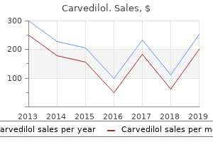
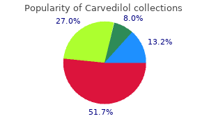
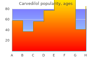
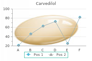
Carvedilol: 25 mg, 12.5 mg, 6.25 mg
Purchase 6.25mg carvedilol fast deliveryProximal stability is important for higher limb operate (Edwards 2002) and conversely instability imposes stresses on the higher limbs during function (Kibler 1998; Magarey & Jones 2003) heart attack belanger remix purchase carvedilol 6.25 mg with amex, which limits their freedom to move away from the body blood pressure medication benefits cheap carvedilol 6.25mg with amex. In a affected person with truncal ataxia blood pressure chart based on age generic carvedilol 6.25mg on line, the upper limbs may be held near heart attack one direction generic 25mg carvedilol mastercard the physique to try and present stability through fixation so that some useful activities may be achieved. These fixation methods, although a solution within the brief time period, might prevent the person exploring their potential for optimising higher limb restoration. The compensatory, extra flexed posture usually related to using walking aids may even cut back the effectivity of balance methods further interfering with upper limb operate. Therefore, there are occasions when you will need to improve walking independence so as to optimise higher limb function. Dynamic stability of the higher and lower trunk, with a steady scapula on the thoracic cage, permits the higher limb to move away from the body, releasing the hand to attain. Hodges (1996) has proven that both decrease limb and trunk muscular tissues fire previous to reaching with the upper limb. Clinically, this is an important consideration as the therapist should not destabilise the patient, by passively taking the upper limb away from the physique, but promote the firing of the postural muscular tissues throughout the trunk for the higher limb to move and be moved away from the body. It appears that deeper trunk muscles activate to stiffen the spine no matter the direction that the upper limb strikes in, but the extra superficial trunk muscles are direction specific (Richardson 2002; Barr et al. The nervous system makes use of these muscle synergies, or patterns of activation, for effectivity (Kavcic et al. Evidence supports the supply of a restraining assist to the trunk which allows for a higher tour of upper limb motion (Michaelsen et al. Thoracic backbone mobility offers a foundation for shoulder activity and is crucial for motion of the upper body and orientation of the higher extremities for use of the arms (Lee et al. The mid-thoracic area of the backbone between T4 and T8 is taken into account to have the best range of rotation (Willems et al. The shoulder complex the shoulder complicated consists of many articulations, muscle tissue, ligaments, bursae and capsules (Mottram 1997; Hess 2000). The glenohumeral joint is the centre of motion at the shoulder advanced (Hess 2000), and it contributes the largest part of the range of movement at the shoulder advanced through its anatomical construction. Efficient neuromuscular exercise especially from the rotator cuff muscular tissues is required for this motion to be managed and to maintain the congruency of the head of humerus within the glenoid fossa. Mottram (1997) describes efficient motion because the integrated and coordinated interaction of the articular, myofascial and neural systems of the physique. The affected person with neurological pathology may have decreased muscular exercise and modifications in sensory and proprioceptive awareness that can impact on the dynamic stability of the shoulder advanced. Glenohumeral stability relies on the place of the scapula on the rib cage, the activity of the supraspinatus muscle and the taut superior facet of the capsule when the higher limb is at relaxation beside the trunk. However, as quickly as the upper limb strikes away from the trunk, more active management is required and the deltoid and 157 Bobath Concept: Theory and Clinical Practice in Neurological Rehabilitation. This highlights the shortage of congruency between the top of humerus and glenoid fossa which can create an inability to selectively activate the rotator cuff musculature. The necessary muscles offering this dynamic stability are subscapularis, supraspinatus, infraspinatus and teres minor (Dark et al. The synchronous contraction of these muscle tissue creates a compressive drive, enabling the humeral head to pivot and glide in the glenoid fossa. Clinically, it is necessary to think about the alignment of the shoulder advanced in the patient who has decreased muscle exercise round this space. Careful positioning and handling of the shoulder complex during each rest and private care duties, corresponding to washing and dressing, helps keep involvement of the higher limb and should forestall trauma to this susceptible space (see Chapter 8). When a affected person is positioned by stabilising the trunk, for instance at rest in aspect lying, the upper limb is allowed to accept the support of the pillow and never the higher limb supporting an unstable trunk. Many authors assist the idea that the scapula position on an upright trunk provides an upward, anterior, lateral-facing glenoid fossa which provides an computerized locking mechanism for the shoulder joint with the upper limb in adduction preventing downward subluxation of the glenohumeral joint (Basmajian 1981; 158 Recovery of Upper Limb Function. The posture of the cervical and thoracic spine has a robust influence on the position and mobility of the scapula and due to this fact the glenohumeral joint (Culham & Peat 1993; Magarey & Jones 2003). The medical implications of decreased antigravity exercise within the trunk embody a loss of scapula alignment and instability of the glenohumeral joint. This additionally applies to positioning the patient within the acute and subacute phases and supporting the hypotonic upper limb, and more importantly, the trunk, with pillows and/or a table to cut back the traction on the delicate tissue and muscles of the upper quadrant. Dysfunction, for example weakness within the scapula musculature, will lead to an alteration in scapula stability leading to shoulder perform turning into less efficient, reducing performance and pre-disposing the individual to injury (Voight & Thomson 2000). Stability at the scapulothoracic joint relies upon not only on the surrounding musculature (Mottram 1997; Voight & Thomson 2000), notably trapezius and serratus anterior, but also on rhomboid major and minor and levator scapulae. These stabilising muscle tissue have to be recruited prior to movement of the upper limb to anchor the scapula (Mottram 1997; Voight & Thomson 2000), and whereas sustaining dynamic stability, they want to additionally provide controlled mobility. A lack of acceptable activation leads to an lack of ability to achieve an efficient reach pattern. However, altering the course of motion could allow for a extra applicable pattern of exercise and is a useful assessment software. The scapula is prepared to transfer in many instructions on the thoracic cage, including elevation, despair, abduction, adduction and rotation (Mottram 1997; Voight & Thomson 2000) and this mobility is essential for: enhancing the congruity of the glenohumeral joint throughout movement; allowing the acromial arch to elevate, so stopping impingement of the humeral tubercles during elevation of the upper limb; a hundred and sixty Recovery of Upper Limb Function. The repeated use of compensatory motion strategies by the patient will have an result on the steadiness of muscle exercise across the shoulder complex, and this will have an impact on functional restoration in the higher limb. This is an space which is particularly troublesome to handle because of the complicated nature of the neurological damage to the methods concerned in postural control and efficient coordination of the patterns of movement needed for higher limb perform. Early work by Inman in 1944 proposed a ratio of two:1 for glenohumeral-to-scapulothoracic movement, that means that when considering 90� elevation of the higher limb, 60� comes from glenohumeral motion and 30� from scapula motion. As talked about previously on this chapter, it is necessary to think about the role of postural stability for mobility and the role of the scapula in achieving range and refinement of motion of the upper limb. Importantly, McQuade and Schmidt (1998) discovered that when the upper limb was loaded, the ratio changed to 4. The thoracic alignment should even be thought of as the scapula should journey across the thoracic cage to allow greater vary of movement within the shoulder advanced. A kyphotic thoracic backbone or broad posterior aspect of the thorax will affect this journey and subsequently the dynamics of scapula stability. This is characterised by force couples of paired muscles that management the motion or position of a joint or body half (Kibler 1998; Voight & Thomson 2000), maintaining maximal congruency between the glenoid fossa and the humeral head. Scapular stabilisation requires a drive couple between the higher and lower portions of trapezius and the rhomboids coupled with serratus anterior, and then as the higher limb is elevated, exercise of the decrease trapezius and serratus anterior muscle tissue is coupled with higher trapezius and rhomboids. Functional reach Although there are events when the higher limb is taken away from the physique with no direct objective of utilizing the hand, for instance to wash beneath your higher limb together with your other hand, many upper limb actions are for the aim of transporting 162 Recovery of Upper Limb Function. When the duty is pointing, all segments of the higher limb are managed as one unit (Shumway-Cook & Woollacott 2007); nevertheless, when the duty is to reach and hold an object, the hand is controlled independently of the opposite upper limb segments. Therefore, reach to grasp may be divided into two parts, the transportation phase and the grasp part. These two parts occur synchronously and appear to be controlled by completely different neural mechanisms.
Syndromes - A fever above 101°F, or your child has a fever above 100.4°F along with diarrhea
- Have you had any injuries
- A piece of tissue or cells are removed and placed on slides.
- Numbness or tingling in the arms and legs
- Testicular cancer (in men)
- Acute trauma of the head and face
Order carvedilol 25mg on lineThe circular limer muscular coat covers the entire size of the colon; the longitudinal coat consists of three bands of muscle tissue heart attack cover purchase 12.5mg carvedilol overnight delivery. The mucous membrane of the colon is epithelial tissue equipped with numerous capillaries for the absorption of water hypertension essential buy carvedilol 6.25mg without prescription. Leukoplakia hyperkeratosis with epithelial atrophy blood pressure chart 13 year old buy carvedilol 25mg on-line, discovered on mucosa of gums arteria lusoria carvedilol 12.5 mg, tongue and inner cheek Mucoepidermoid carcinoma salivary gland neoplasm, contains plenty of mucus. Squamous cell carcinoma invades domestically and metastasizes to lymph nodes, frequent in lips, tongue and oral cavity. Stomach: Adenocarcinoma stomach often presents as an ulcer with a hard, rolled, agency white edge - infiltrates could involve the muscle wall. Gastric polyp giant projection overgrown with epithelium organized round stalk which carries blood vessels and lymphatics - could additionally be premalignant. Pectic ulcer a localized ulcer of the visceral mucosa which may cause bleeding (haematemesis) or perforation. Pancreas: Adenocarcinoma pancreas a mucus-producing tumour typically with recognizable glands. Cystic fibrosis generalized abnormality of the secreting glands such as people who manufacture mucus and sweat. The mucus is thick and sticky and the sweat glands have a excessive focus of salt. When the lungs are affected, bronchitis occurs, resulting in persistent cough, wheezing, dyspnoea, and emphysema. Diabetes a disease where the islets of Langerhans within the pancreas fail to produce adequate insulin. Liver and gallbladder: Acute catarrhal jaundice synonymous with infectious hepatitis. Medical Terminology Course 35 Cholelithiasis presence of stones in the gallbladder. Cirrhosis of liver fibrosis of the liver, Hepatoma major tumour of the liver, Hepatitis irritation of the liver. Jaundice yellow pores and skin and sclera because of liver cell changes and obstruction causing bile pigment, bilirubin, to be diffused into the blood. Enteritis any inflammatory situation of the small intestine, Idiopathic sprue atrophy of the intestinal vil, often in the jejunum, with thinning mucosa causes malabsorption syndrome. Obstruction of the gut causes distention of the visais above the lesion and collapse distal to the lesion. Parasitic ailments common infestations such because the pinworm, Trichinella spiralis (from pork), beef tapeworms (Taenia saginata) or pork tapeworm (Taenia solium). Peritonitis irritation of the peritoneum could additionally be caused by rupture of an stomach organ. Volvulus a twisting of the mesentery inflicting obstruction of the blood supply and mechanical blockage of the lumen of the gut. Large gut: Adenocarcinoma of colon epithelial in origin, involving the caecum, sigmoid and rectum predominantly. Carcinoid tumour of appendix normally benign - adenocarcinoma of the appendix is uncommon, colloid in nature. If it ruptures, the peritoneal cavity is filled with jelly-like substance (pseudomyxorna peiitonei). Diverticulosis outpouching of mucosa from the lumen as a result of defective musculature (diverticulitis) with subsequent inflammation. Familial polyposis massive numbers of polyps all through the lumen of the colon, premalignant in nature, genetic in origin. Ulcerative colitis an ulcerative illness of the colon characterised by violent diarrhoeic episodes with blood and mucus in the watery stool - a premalignant condition. Mesentery: Fat necrosis benign inflammatory situation - small quite a few white lesions in mesentery. Panniculitis (L pannus = cloth) inflammation of the fatty portion of the panniculus adiposus, the superficial fasciae with fat in its areolar substance. Each kidney is situated laterally to the spinal column, within the upper part of the abdomen. They are retroperitoneal, embedded in a mass of fatty tissue, which is surrounded by a fibrous covering known as renal fascia. The renal artery which branches from the aorta after a number of subdivisions (Diagram 16) finish in a cluster of capillaries to form the glomerulus. The glomerulus is surrounded by a dosed finish of a protracted tortuous renal tubule, the nephron. Blood leaving the glomerulus flows to a secondary capillary network around the tubules of its personal nephron earlier than draining into a vein. Formation of urine Through the capillaries of the glomeruli roughly 120 ml of water and salts are filtered from the blood each minute. Cells and plasma proteins are too large to cross by way of the capillary membranes into the renal tubule in a healthy unless their concentration within the physique is too high. Additional water is reabsorbed within the distal convoluted tubule and partly in the accumulating tubules. These muscular tubes are about ten inches (25 cm) in size, starting on the renal pelvis. The wall of the ureter has three layers, an outer fibrous coat, a center easy muscle layer which propels urine alongside the ureter and an inner mucous membrane. The trigone is smooth, even when the bladder is empty and the remainder of the smooth muscle is in folds. The bladder has three layers, aside from the superior and posterior features which are coated by peritoneum. These are a fibrous outer layer, a smooth muscle layer and a mucous membrane lining the cavity. The proximal finish consists of a round easy muscle known as the inner sphincter. The exterior sphincter is a circular striated muscle which is beneath voluntary management. The filtrate accommodates glucose, salt, urea, uric acid, potassium, phosphates, sulfates, and so forth. However, the body must retain certain of these substances for fluid and electrolyte balance. Thus, because the filtrate passes along the tubule of the nephron, the filtrate is concentrated and essential substances are returned to the circulation via the second capillary community which surrounds the tubule. Essential or "high threshold" substances are additionally reabsorbed here, together with glucose, sodium chloride and amino acids, Female urethra the feminine urethra is roughly 4-5 an, 1. Ammonia as sodium is removed from the filtrate again into the blood stream ammonia is formed.
Best 6.25mg carvedilolThe nerve move through the posterior andmiddle cranial fossae and divides into superior and inferior divisions close to the superior orbital fissure blood pressure ranges by age and gender order carvedilol 6.25 mg without prescription. The trochlear nerve this is the somatic motor nerve a hundred and seventy Human Anatomy and Physiology supply to the superior indirect blood pressure stroke level cheap carvedilol 6.25mg on-line. The fibres move posteriorly and bear a dorsal decussation with the nerve of the other side caudal to the inferior colliculi the nerve then passes forwards via the posterior and center cranial fossae pulse pressure 58 cheap carvedilol 25mg without a prescription,enters the orbit through the superior orbital fissure and provides superior indirect blood pressure medication bananas buy carvedilol 6.25mg free shipping. The third branch is joined by motor fibers to the muscular tissues of mastication (chewing). The nerve leaves the inferior border of the pons near the midline, passes forwards by way of the posterior and middle cranial fossae, the cavernous sinus and the orbit, and provides lateral rectus. The vestibulocochlear nerve accommodates special sensory fibers for hearing as properly as these for stability from the semi circular canals of the internal ear. The glossopharyngeal nerve contains basic sensory fibers from the back of the tongue and the pharynx (throat). This nerve additionally incorporates sensory fibers for style from the posterior third of the tongue, secretary fibers that supply the most important salivary gland (parotid), and motor nerve fibers to control the swallowing muscle tissue within the pharynx. This nerve also incorporates motor fibers to the larynx (voice box) and pharynx, and to glands that produce digestive juices and different secretions. The accent nerve (formerly known as the spinal accessory nerve) is a motor nerve with two branches. The hypoglossal nerve, the last of the 12 cranial nerves, carries impulses controlling the muscular tissues of the tongue. The roots are fashioned from a number of rootlets which emerge from the anterolateral and posterolateral sulci of the spinal cord. The ventral root carries efferent (motor) fibres from the cord and the dorsal root, afferent (sensory) fibres to the wire. The cell our bodies of the sensory fibres are situated in a ganglion on the dorsal root. Each nerve leaves the vertebral canal by way of an intervertebral foramen and soon divides into a big ventral and smaller dorsal ramus (branch). The adjoining ventral rami of most areas communicate to type plexuses (cervical, brachial and lumbosacral) while these of the thoracic area turn out to be the intercostals and subcostal nerves. The dorsal rami pass backwards into the postvertebral muscle tissue and divide into medial and lateral branches. These rami provide the muscular tissues and skin over the posterior aspect of the body but give no branches to the limbs. The ventral rami 173 Human Anatomy and Physiology supply the anterior and lateral wall of the again and the lower limbs. Branches of the Spinal Nerves Each spinal nerve continues solely a very brief distance away from the spinal twine and then branches into small posterior divisions and quite giant anterior divisions. The larger anterior branches interlace to kind networks called plexuses, which then distribute branches to the body components. The cervical plexuses supplies motor impulses to the muscles of the neck and receive sensory impulses from the neck and the again of the pinnacle. The brachial plexus sends numerous branches to the shoulder, arm, forearm, wrist, and hand. The largest of these branches is the sciatic nerve, which leaves the dorsal part of the pelvis, passes beneath the gluteus maximus muscle, and extends down the back of the thigh. These afferent impulses from the viscera are translated into reflex responses with out reaching the upper heart of the mind; the sensory neurons from the organs are grouped with those who come from the pores and skin and voluntary muscle tissue. In distinction, the efferent neurons, which provide the glands and the involuntary muscular tissues, are organized very in a unique way from those who supply the voluntary muscle tissue. In these g~ every message is transferred at a synapse from the first neuron to a second one and from there to the muscle or gland cell. This differs from the voluntary (somatic nervous system, in which each motor 176 Human Anatomy and Physiology nerve fiber extends all the means in which from the spinal cord to the skeletal muscle with no intervening synapse. Some of the autonomic fibers are within the spinal nerves; some are within the cranial nerves. The sympathetic pathways begin within the spinal twine with cell bodies in the thoracic and lumbar areas, the thoracolumbar area. The sympathetic fibers come up from the spinal cord on the stage of the primary thoracic nerve right down to the level of the second lumbar spinal nerve. From this a half of the twine, nerve fibers extend to ganglia the place they synapse with a second set of neurons, the fibers of which lengthen to the glands and involuntary muscle tissues. Many of the sympathetic ganglia type the sympathetic chains, two twine like strands of ganglia that reach alongside both facet of the spinal column from the lower neck to the upper stomach region. The nerves that supply the organs of the belly and pelvic cavities synapse in three single ganglia farther from the spinal wire. The second neurons of the sympathetic nervous system act on the effectors by releasing the neurotransmitter epinephrine adrenaline. This system is therefore described as adrenergic, which implies "activated by 177 Human Anatomy and Physiology 2. The parasympathetic pathways start in the craniosacral areas, with fibers arising from cell our bodies of the midbrain, medulla, and decrease (sacral) part of the spinal cord. From these facilities the primary set of fibers extends to autonomic ganglia which are often located close to or within the partitions of the effector organs. The pathways then continue along a second set of neurons that stimulate the involuntary tissues. These neurons launch the neuro transmitter acetylcholine, leading to the outline of this method as cholinergic (activated by acetylcholine). These actions are all carried on mechanically; whenever any changes happen that decision for a regulatory adjustment, the adjustment is made with out acutely aware awareness. The sympathetic part of the autonomic nervous system tends to act as an accelerator for these organs needed to meet a annoying state of affairs. This produces hormones, together with epinephrine, that put together the body to meet emergency conditions in some ways. Increase in blood strain due partly to the more practical heartbeat and partly to constriction of small arteries in the skin and the inner organs 5. Dilation of blood vessels to skeletal muscles, bringing more blood to these tissues 179 Human Anatomy and Physiology 6. The sympathetic system additionally acts as a brake on those methods not directly involved within the response to stress, such because the urinary and digestive methods. Under these circumstances, when food does reach the stomach, it appears to stay there longer than usual. The parasympathetic a part of the autonomic nervous system nonnal1y acts as a balance for the sympathetic system once a crisis has passed. The parasympathetic system brings about constriction of the pupils, slowing of the center price, and constriction of the bronchial tubes. It also stimulates the formation and launch of urine and exercise of the digestive tract. Saliva, for example, flows more simply and profusely and its amount and fluidity enhance.
Trusted carvedilol 25mgThromboplastin reacts with certain protein factors and calcium ions to type prothrombin activator pulse pressure 40 generic 6.25 mg carvedilol, which in flip reacts with calcium ions to convert the prothrombin to thrombin blood pressure chart range order carvedilol 12.5 mg without prescription. Fibrin forms a network of threads that entraps red blood cells and platelets to kind clot blood pressure lowering purchase 6.25mg carvedilol with amex. Thromboplastin Ca++ Prothrombin Thrombin Fibrinogen Fibrin threads + Blood cells and plasma Clot Blood Typing and Transfusions Blood Groups If for some cause the quantity of blood in the physique is severely decreased arrhythmia specialists purchase 12.5 mg carvedilol fast delivery, by way of haemorrhage or illness, the body cells endure from lack of oxygen and food. The obvious measure to absorb such an emergency is to inject blood from one other individual into the veins of the patient, a procedure called transfusion. These reactions are decided largely by certain proteins, known as antigens, on the surface membrane of the purple blood cells. There are many kinds of these proteins however solely two groups are notably prone to cause a transfusion response, the so-called A and B antigens and the Rh issue. Blood serum containing antibodies that may agglutinate and destroy purple cells which have A antigens on the surface is called anti-A serum; blood serum containing antibodies that can destroy red cells with B antigen on the floor known as anti-B serum. However, due to other elements that might be present within the blood, willpower of blood type have to be accompanied by further exams (cross matching) for compatibility earlier than a transfusion is given. The Rh issue Rh issue is another pink cell antigen that determines the blood group. Those individuals who possess this antigen of their red cell floor are said to be Rh constructive. If Rh optimistic blood is given to an Rh negative particular person, she or he could turn out to be sensitized to the protein within the Rh positive blood. This 257 Human Anatomy and Physiology condition is called erythroblastosis fetalis, or haemolytic illness of the new child. Slightly greater than a fist, this organ is situated between the lungs in the heart and a bit to the left on the midline of the physique. The proven truth that its fee of beating is affected by the feelings may be liable for the very frequent references to the heart in song and poetry. However, the very important features of the heart and its issues are of extra practical significance to us. The endocardium is a really skinny smooth layer of cells that resembles squamous epithelium. The epicardium types the skinny outermost layer of the center wall and is continuous with the serous lining of the fibrous sac that encloses the guts. The serous lining of the pericardial sac is separated from the epicardium on the heart surface by a skinny fluid- crammed area. Two Hearts and a Partition Physicians usually check with the best heart and the left coronary heart. The two sides are completely separated from each other by a partition referred to as the septum. The higher part of this partition known as interartrial septum; whereas the bigger the lower portion is called interventricular septum. The proper atrium is a thin-walled chamber that receives the blood retuning from the body tissues. This blood, which is low in oxygen, is carried within the veins, the blood vessels leading to the center from the body tissues. The proper ventricle pumps the venous blood received from the right atrium and sends it to the lungs. The left atrium receives blood high in oxygen content material as it returns from the lungs. The left ventricle, which has the thickest partitions of all, pumps, oxygenated blood to all elements of the physique. This blood goes through the arteries, the vessels that take blood from the guts to the tissues. Four Valves Since the ventricles are the pumping chambers, the valves, that are all a method, are positioned on the entrance and the exit of every ventricle. The entrances valves are the atrioventricular valves, whereas the exit valves are the semilunar valves. The proper atrioventricular valve also is called the tricuspid valve, because it has three cusps, or flaps, that open and closes. When this valve is open, blood flows freely from the proper atrium into the best ventricle. It has two somewhat heavy cusps that permit blood to move freely from the left 261 Human Anatomy and Physiology atrium into the left ventricle. However, the cusps shut when the left ventricle begins to contract; this prevents blood from returning to the left atrium and ensures the ahead circulate of blood into the aorta. Both the tricuspid and mitral valves are attached by the use of thin fibrous threads to the wall of the ventricles. The pulmonic (semilunar) valve is positioned between the proper ventricle and the pulmonary artery that results in the lungs. As quickly as the proper ventricle has completed emptying itself, the valve closes in order to prevent blood on its approach to the lungs from returning to the ventricle. Following contraction of the left ventricle, the aortic valve closes to stop the move of blood again from the aorta to the ventricle. Blood Supply to the Myocardium Although blood flows by way of the heart chambers, only the endocardium comes into contact with it. Therefore, the myocardium must have its own blood vessels to present oxygen and nourishment and to remove waste merchandise. After passing via capillaries in the myocardium, blood drains into the cardiac veins and eventually into the coronary (venous) sinus for return to the right atrium. Valves of the guts, seen from above, within the closed place (From Memmler and Wood: the Human Body in Health and Disease, ed 6, Philadelphia, 1987, J. The blood is squeezed by way of the chambers by a contraction of coronary heart muscle starting within the thin-walled higher chambers, the atria, adopted by a contraction of the thick muscle of the decrease chambers, the ventricles. The contraction of the walls of the atria is completed on the time the contraction of the ventricles begins. Thus, the resting section (diastole) begins in the atria simultaneously the contraction (systole) begins in the ventricles. After the ventricles have emptied, both chambers are relaxed for a brief period of time as they fill with blood. The fibers are interwoven so the stimulation that causes the contraction of one fiber leads to the contraction of the entire group. This performs an essential position in the strategy of conduction and the working of the guts muscle. When the heart chamber is stuffed and the wall stretched (within limits), the contraction is strong. As extra blood enters the heart, as occurs throughout train, the muscle contracts, with greater energy so push the bigger volume of blood out into the blood vessels. The volume of blood pumped by each ventricle in 1 minute is termed the cardiac output. It is set by the quantity of blood ejected from the ventricle with each beat-the stroke 265 Human Anatomy and Physiology volume-and the number of beats of the heart per minute-the heart rate.
Buy cheap carvedilol 25mg onlineAt the higher end of the larynx are the vocal cords pulse pressure values order carvedilol 6.25mg otc, which serve within the manufacturing of speech arteria inominada cheap carvedilol 25mg on-line. The nasal cavities arteria obturatriz carvedilol 6.25mg low cost, the sinuses hypertension epidemiology buy carvedilol 25 mg with visa, and the pharynx all serve as resonating chambers for speech, simply as the cupboard does for a stereo speaker. The space between these two vocal cords is recognized as the glottis, and the little leaf-shaped cartilage that covers the larynx during swallowing is called the epiglottis. As the larynx moves upward and ahead during swallowing, the epiglottis strikes downward, overlaying the opening into the 297 Human Anatomy and Physiology larynx. You can feel the larynx move upward toward the epiglottis throughout this process by inserting the flat ends of your fingers in your larynx as you swallow. The cilia lure mud and other particles, transferring them upward to the pharynx to be expelled by coughing, sneezing, or blowing the nostril. The Trachea (Windpipe) the trachea is a tube that extends from the lower edge of the larynx to the higher part of the chest above the heart. These cartilages, shaped somewhat like a tiny horseshoe or the letter C, are discovered along the entire size of the trachea. All the open sections of those cartilages are at the back so that the esophagus can bulge into this part throughout swallowing. The Bronchi and Bronchioles the trachea divides into two bronchi which enter the lungs. The proper bronchus is significantly larger in diameter than the left and extends downward in a more vertical course. Each bronchus enters the lung at a notch or 298 Human Anatomy and Physiology depression referred to as the hilus or hilum. The Lungs the lungs are the organs by which exterior respiration takes place by way of the extremely skinny and delicate lung tissues. The two lungs, set aspect by facet within the thoracic cavity, are constructed in the following manner: Each bronchus enters the lung on the hilus and immediately subdivides. The bronchi subdivide repeatedly, forming progressively smaller divisions, the smallest of which are called bronchioles. At the tip of every of the smallest subdivisions of the bronchial tree, referred to as terminal bronchioles, is a cluster of air sacs, resembling a bunch of grapes. This very thin wall provides easy passage for the gases getting into and leaving the blood as it circulates by way of hundreds of thousands of tiny capillaries of the alveoli. Certain cells within the alveolar wall produce surfactant, a substance that prevents the alveoli from collapsing by decreasing the floor tension ("pull") of the fluids that line them. Because of the numerous air spaces, the lung is gentle in weight; usually a piece of lung tissue dropped into a glass of water will float. In the lungs bl9od passes via the capillaries across the alveoli, the place the fuel exchange takes place. The pleural cavity across the lungs is an air-tight house with a partial vacuum, which causes the strain on this area to be lower than atmospheric strain. Because the pressure inside the lungs is greater than that within the surrounding pleural cavity, the lungs are likely to remain inflated. The complete thoracic cavity is versatile, able to expanding and contracting along with the lungs. The region between the lungs, the mediastinum, incorporates the center, nice blood vessels, esophagus, trachea, and lymph nodes. Physiology of Respiration Pulmonary Ventilation Ventilation is the motion of air into and out of the lungs, as in respiratory. The diaphragm is a strong dome-shaped muscle hooked up around the base of the rib cage. The contraction and rest of the diaphragm trigger a piston-like downward movement that lead to a rise within the vertical dimension of the chest. The rib cage can additionally be moved upward and outward by contraction of the external intercostals muscular tissues and, throughout exertion, by contraction of different muscle tissue of the neck and chest. During quiet respiratory, the motion of the diaphragm accounts for a lot of the increase in thoracic volume. When the strain drops to slightly under atmospheric stress, air is drawn into the lungs. In exhalation, the passive section of breathing, the muscular tissues of respiration chill out, permitting the ribs and diaphragm to return to their unique positions. During compelled exhalation, the internal intercostals muscles and the muscle tissue of the belly wall contracts, pulling the underside of the rib cage in and down. Air Movement Air enters the respiratory passages and flows by way of the ever-dividing tubes of the bronchial tree. Here the air strikes by diffusion, which quickly equalizes any variations within the quantities of gases current. Each breath causes comparatively little change within the gasoline composition of the alveoli, but normal steady respiration ensures the presence of adequate oxygen and the removing of carbon dioxide. Table 17-1 Breathing Volumes Volume Tidal quantity Definition the amount of air moved into or out of the lungs in quiet, relaxed respiration Average value 500 cc Vital capacity the quantity of air that can be expelled from the lungs by most exhalation inhalation following most 4800 cc Residual volume Total capacity Functional residual capacity lung the amount of air that continues to be in the lungs after most exhalation the whole quantity of air that can be contained within the lungs after maximum inhalation the quantity of air remaining within the lungs after normal exhalation 1200 cc 6000 cc 2400 cc Regulation of respiration Regulation of respiration is a fancy process that should hold tempo with moment-to-moment modifications in cellular oxygen necessities and carbon dioxide manufacturing. Regulation 305 Human Anatomy and Physiology depends primarily on the respiratory management facilities located within the medulla and pons of the mind stem. Respiration is regulated so that the degrees of oxygen, corbon dioxide, and acid are saved inside sure limits. From the respiratory center within the medulla, motor nerve fibers lengthen into the spinal wire. From the cervical (neck) a half of the wire, these nerve fibers continue via the phrenic nerve to the diaphragm. The diaphragm and the opposite muscle tissue of respiration are voluntary in the sense that they are often regulated by messages from the upper mind centers, notably the cortex. It is feasible for a person to intentionally breath more rapidly or more slowly or to hold his breath and never breath in any respect for a time. Usually we breath without thinking about it, whereas the respiratory centers within the medulla and pons do the controlling. These receptors are present in structures referred to as the carotid and aortic our bodies, as nicely as out facet the medulla of the mind stem. The carotid our bodies are situated close to the bifurcation of the frequent carotid arteries, whereas the aortic bodies are located within the aortic arch. These bodies comprise many small blood vessels and sensory neurons, that are 306 Human Anatomy and Physiology sensitive to decreases in oxygen supply as nicely as to will increase in carbon dioxide and acidity (H+). Impulses are despatched to the brain from the receptors in the carotid and aortic our bodies. It should first be broken down into particles small enough to cross through the cell membrane. After digestion, food must be carried to the cells in each a part of the physique by the circulation.
Buy generic carvedilol 25mgThis was combined with facilitation of ahead weight switch over the foot in criminal mendacity as a basis for selective 107 Bobath Concept: Theory and Clinical Practice in Neurological Rehabilitation heart attack diet buy cheap carvedilol 25 mg. Increased stability on the pelvis allowed him to improve his control in forward translation of the knee blood pressure medication depression carvedilol 6.25mg for sale, an important element in environment friendly movement from standing to sitting hypertension lab tests generic carvedilol 25mg on line. This element was additionally practised within the context of standing hypertension over 65 buy carvedilol 12.5mg cheap, using the wall as an environmental support. Summary points from the medical instance the early hypotonic patient supplies a problem in rehabilitation. Minimising the learning of inefficient compensation and but maximising independence is a major goal. Systematic evaluation and specific intervention to influence orientation, postural stability and activation allows optimum performance as a basis for continued development in course of practical independence. Part task and whole task practice in a wide selection of settings will assist transferability of the skill. Achieving the appropriate alignment and exercise of all physique segments is necessary each prior to and through execution of the switch. A sturdy relationship between sensory enter and motor output exists, for example the heels actively moving all the method down to the floor is facilitatory to activation of the decrease limb extensor musculature, optimising a extra automatic drive to increase the physique. Independent standing and early facilitated stepping for switch with elevated confidence and stability. Key Learning Points Acquiring independence in shifting between sitting and standing is important for achieving unbiased mobility. The extensive body of literature obtainable offers an overview of the weather of the transfer, however to apply this information in a scientific setting the limitations imposed by the research methodology should be thought of. A clear understanding of the interplay between postural stability and selective movement is required to guide scientific reasoning and intervention. Achieving functional independence on the earliest alternative is a key goal of rehabilitation but have to be combined with the relearning of acceptable motion components if continued recovery is to be optimised and secondary adaptive modifications minimised. As can be seen in the medical instance, many different factors could need to be considered in developing an individualised treatment intervention to optimise their potential in any respect stages of rehabilitation. The Journals of Gerontology Series A: Biological Sciences and Medical Sciences, 60A, 1546�1552. The Control of Locomotion Ann Holland and Mary Lynch-Ellerington Introduction Walking is commonly one of the most necessary goals for patients with neurological circumstances taking part in rehabilitation (Mudge & Stott 2007). This chapter will consider key elements of locomotion and the medical software. The specific goals of the chapter are to: introduce key features of bipedalism; explore particular options of motor management; think about gait initiation; highlight scientific problems and interventions that can be used in the hemiparetic population; adapt medical interventions for persons with other neurological conditions. Key aspects of bipedalism Human erect locomotion is exclusive amongst residing primates and demonstrates particular biomechanical options that make it mechanically efficient and enduring (Lovejoy 2004). The regulation of bipedal stance and gait requires particular neuronal mechanisms to maintain the body in an upright position (Dietz & Duysens 2000). Humans have developed an upright stance capable of endurance walking over very long distances. These options, in accordance with the laws of kind and performance, are neuroplastically matched by the motor patterns generated in the nervous system (Grasso et al. Man is capable of locomotion over a variety of velocities, from very sluggish speeds to short time efficiency at speeds up to and above 10 metres per second 117 Bobath Concept: Theory and Clinical Practice in Neurological Rehabilitation (Neptune & Sasaki 2005). Key evolutionary elements underpinning bipedalism (Lovejoy 2004) are: the unique human abductor apparatus providing pelvic stabilisation throughout single leg stance; the development of a lordosis and the repositioning of the centre of mass; the expanded position of gluteus maximus from which to control trunk extension at heel strike. Proper execution of locomotion requires integration of neuronal subsystems involved in the creation of postural and locomotor management (Mori et al. The evidence now strongly helps the idea that the trunk is an energetic component of postural management preceding the initiation of strolling and not a passenger as may have originally been thought (Perry 1992). Locomotion must assure a ahead progression suitable with dynamic equilibrium, adapting to doubtlessly destabilising elements in an anticipatory trend by the use of coordinated synergies of the upper limbs, trunk and decrease limbs (Grasso et al. Integrated control of posture and strolling is made potential as a end result of these two motor functions share some common organisational principles. Firstly, the frame of reference for the kinematic coordination for each postural responses and locomotion seems to be anchored to the vertical. Secondly, management of the place of the centre of mass for static or dynamic equilibrium is concerned in each gait and posture (Grasso et al. The concept of built-in management of posture and locomotion is central to the clinical apply of the Bobath Concept. This stems from neurophysiological evidence with respect to nervous system control and its relationship with afferent information updating physique schema. Neurophysiological research indicate that the management of posture and locomotion are interdependent, and interdependency exists at many various ranges within the nervous system (Patla 1996). Essential requirements for locomotion Walking is a sophisticated motor act requiring the coordination of trunk and limb muscle tissue crossing many joints (Mackay-Lyons 2002). It is a fundamental prerequisite of daily life in addition to some of the automatic, and is the useful result of the interplay of biomechanical, neurophysiological and motor management systems. The want to regain strolling capability after neurological dysfunction is often the primary goal of rehabilitation, and as a consequence a lot time and energy is dedicated to its retraining. Successful human locomotion is characterised by a fundamental locomotor pattern which strikes the body within the desired course and postural management to support the physique towards gravity (Shumway-Cook & Woollacott 2007). Walking should also be adaptable to meet the needs of the individual and the calls for of the surroundings. This is achieved by way of the regulation of postural tone, significantly in the extensor antigravity musculature, and correct foot placement (Grillner et al. In order for these task requirements to be met, a non-hierarchical tripartite management system is required. Supraspinal and sensory influences are extremely highly effective and facilitate the power to modify limb movements whereas ensuring the upkeep of balance and posture (Sorensen et al. The cortical management of walking is advanced with respect to the relationship of cortical and subcortical constructions involved. For walking to be actually functional, it has to be of affordable velocity and distance, for example to permit crossing the street in a given time at a pedestrian crossing. In terms of home walking, the minimal distance required to walk may be from the sitting room to the bathroom (Bohannon 2001). Walking in a easy surroundings of open space is usually difficult for patients, and walking within the advanced setting of a busy avenue or buying centre could additionally be impossible with out the element of automaticity. Taking the patient to a dual tasking degree is a vital role of rehabilitation, as a end result of it represents life in the real world. Feedback through spinoreticular neurons and inputs from different areas of the mind seem to be essential to stabilise the locomotor rhythm (Mackay-Lyons 2002). Clinical relevance Initiation of step one from a quiet stance entails moving the centre of mass outdoors the bottom of assist, transferring weight over the help limb and moving the swing limb ahead (Patla 1996). Corticospinal drive is involved in the initiation of the gait cycle by way of flexion of the swing leg in direction of the first heel strike.
References - Koppie TM, Vickers AJ, Vora K, et al: Standardization of pelvic lymphadenectomy performed at radical cystectomy: can we establish a minimum number of lymph nodes that should be removed?, Cancer 107:2368n2374, 2006.
- Tiggemann M. Media influence on body image development. In: Cash TF, Pruzinky T, eds. Body Image: A Handbook of Theory, Research and Clinical Practice. New York: The Guilford Press, 2002: 91-8.
- Pfeiffer RA. Dominant Erbliche Akrocephalosyndaktylie. Z Kinderheilkd 1964;90:301-320.
- Cartledge, J.J., Cross, W.R., Lloyd, S.N., Joyce, A.D. The efficacy of a range of contact media as coupling agents in extracorporeal shockwave lithotripsy. BJU Int 2001;88: 321-324.
- Balling G, Vogt M, Kaemmerer H, et al. Intracardiac thrombus formation after the Fontan operation. 20.
- Steed DL, Brown B, Reilly JJ, et al. General surgical complications in heart and heart-lung transplantation. Surgery. 1985;98:739-745.
- Bohrod MG. The pathogenesis of acute appendicitis. Am J Clin Pathol 1946;16:752.
|

