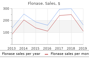
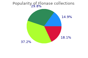
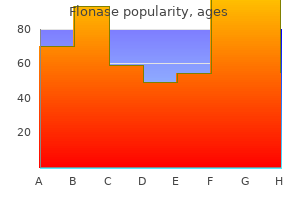
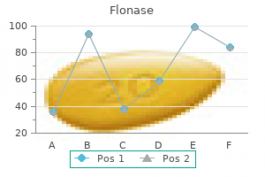
Flonase
Caleb P. Bupp, M.D. - Department of Medical Genetics
- Spectrum Health System
- Grand Rapids, Michigan
Trusted 50mcg flonaseElectron microscopy the ultrastructural options are completely different within the various sorts allergy ready buy 50 mcg flonase. In the grownup kind allergy testing methods cheap flonase 50 mcg otc, there are large quantities of amorphous and granular material with some wavy allergy medicine that starts with a c buy flonase 50mcg low price, ill-defined allergy testing kits discount flonase 50 mcg fast delivery, brief, and branching filaments. In the juvenile type, there are fibrillary plenty with some whorling, uncommon nuclear remnants, and some melanosomes and desmosomes. The deposits extend extra deeply than in colloid milium, and clefting is usually much less conspicuous. Therefore, in the latter situations, an association with elastic fibers is often noticed outright or can be inferred, with both H&E-stained sections and in tissues stained with the Verhoeff�van Gieson methodology or other elastin stains, and the colloid materials is adverse for keratins. As beforehand famous, keratin positivity additionally characterizes the deposits in macular and lichenoid amyloidosis. However, stains for amyloid are often weak or adverse in juvenile colloid milium. Although weak Congo purple positivity has been described, immunostaining for amyloid P substance is unfavorable. Crystal-shaped empty spaces could additionally be seen inside the material, and occasionally birefringent crystals have been present. It is characterized by amorphous, eosinophilic materials within and around blood vessels related to acute or persistent irritation. Small spindled cells, a few of that are surrounded by clear areas, are recognized inside a hyaline eosinophilic matrix. The time period has been applied to a heterogeneous group of deposits that embody amyloid, colloid bodies, Russell our bodies, and elastic globes. Colloid our bodies are derived from degenerate keratinocytes, often associated with the lichenoid reaction pattern. Elastic globes had been first described within the 19th century and had been for a time considered a diagnostic sign in cutaneous lupus erythematosus or scleroderma. It has been advised that elastic globes in some circumstances could represent the top stage of degenerated colloid our bodies; this has not been confirmed by immunohistochemistry, which reveals that elastic globes have an immunological profile just like that of elastic fiber microfibrils. For comfort, the varied heavy metals and drugs that produce deposits and/or pigmentary changes are discussed right here as well. For completeness, observe that the shaving heads and foils utilized in electrical shavers may be a day by day source of iron�chromium�nickel contamination of the skin, main theoretically to allergic reactions. Pigment granules are additionally found within the endothelial cells of blood vessels and the basement membrane of sweat glands. Rarely, international body large cells surround the fibers, but this happens extra usually in extracutaneous sites. The term ochronosis can be used for the deposition of similar hydroquinone derivatives in sure exogenously induced situations that generally followed the topical use of phenol within the treatment of leg ulcers and of picric acid in the treatment of burns (both procedures have now been abandoned) and which are nonetheless seen as a complication of the oral administration668,669 or intramuscular injection670 of antimalarial medication and the topical use of hydroquinone bleaching creams in black 446 Section4 � TheDermisandSubcutis in macrophages in a perivascular place and round appendages. The pigment in the antimalarial-induced instances usually stains positively for melanin and hemosiderin,668 whereas within the different varieties the fibers and smaller deposits are normally unfavorable with these stains and likewise with elastic tissue stains. Black pigment is seen in macrophages and lying free in a predominantly perivascular location. In one telephone study reported from the United States, 24% of respondents had tattoos,698,699 whereas an nearly identical incidence was obtained in a survey of university undergraduates. The infections reported have included pyogenic infections, syphilis, leprosy, tuberculosis,710 tetanus, chancroid, verruca vulgaris,706,711�714 vaccinia, herpes simplex and zoster, molluscum contagiosum,715 viral hepatitis, and a dermatophyte an infection. A overseas physique granulomatous reaction has not been recorded besides within the presence of different severe reactions. Recently, attempts have been made to correlate the ultrastructural options with the pigment used, as decided by X-ray microanalysis methods. The mechanism by which the heavy metals stimulate melanin production is uncertain. Histopathology767,771 There could also be some thinning of the dermis and increased melanin pigment within the basal layer. Golden brown granules of hemosiderin are present in the basement membrane area of the sweat glands and in macrophages within the free connective tissue stroma of these glands. A small amount can usually be seen associated with sebaceous glands and their stroma. The bronze shade outcomes from elevated melanin in the basal layers of the dermis and, to a lesser extent, some coexisting thinning of the dermis. There are fine silver particles alongside the connective tissue sheath of the hair follicle (arrow). Localized green discoloration of the palms and soles has been reported in an grownup with hyperbilirubinemia. Localized argyria has been reported after extended topical exposure,800,801 on the website of implanted acupuncture needles,802�804 and from the wearing of silver earrings in pierced ears. Histopathology784 There are a number of, minute, brown-black granules deposited in a bandlike method in relation to the basement membranes of sweat glands. Scanning electron microscopy has proven that the granules are bigger and more abundant in uncovered than nonexposed pores and skin. Dermoscopy of argyria reveals blue-gray dots, annular buildings, and streaks (silver deposits) throughout a yellow background (the grenz zone of uninvolved papillary dermal collagen). This differs from the homogeneous blue areas, globules, or red pigment or streaks seen in melanocytic lesions. The accidental or deliberate implantation of mercury into the pores and skin ends in a granulomatous foreign physique big cell response. Differential analysis of argyria, chrysiasis, and hydrargyriasis the traits of the granules in these conditions distinguish the pigmentation they produce from that due purely to melanin or to pigment-producing medication similar to minocycline, phenothiazines, or antimalarials, which may variably embrace melanin, a melanin-like drug metabolite, or iron. With light microscopy, it may be difficult to distinguish among gold, silver, and mercury deposits. However, gold granules are larger and more irregular than those of silver and are found predominantly within macrophages. Silver particles are largely extracellular and tend to be deposited on basement membranes, round vessels, and on elastic fibers. Mercury granules are also found most commonly in macrophages or occasionally in the epidermal basilar layer. The orange-red birefringence of gold particles with cross-polarized mild may be a novel function. Further distinction could be made with ultrastructural research, based mostly on not solely the distribution but additionally the relative dimension of the granules (gold is larger than silver; silver is larger than mercury) and their aggregation properties. If needed, X-ray microanalysis and related strategies can present definitive identification of the material. Histopathology784,816,822 Small round or oval black granules, irregular in dimension, are current in dermal macrophages that are inclined to localize around blood vessels in the upper and mid dermis. Electron microscopy Electron microscopy exhibits electron-dense particles in phagolysosomes of macrophages. Cutaneous nodules containing globules of mercury have been reported in one patient, as a reaction to oral mercury.
Purchase 50mcg flonase with visaClear cell sarcoma (malignant melanoma) of soppy components: A clinicopathologic examine of 30 circumstances allergy medicine vegan cheap 50 mcg flonase overnight delivery. Translocation t(12;22)(q13;q13) is a nonrandom rearrangement in clear cell sarcoma allergy medicine non antihistamine generic 50mcg flonase fast delivery. Diagnosis of clear cell sarcoma by real-time allergy forecast livermore ca buy 50mcg flonase with amex, reverse transcriptase-polymerase chain response analysis of paraffin embedded tissues: Clinicopathologic and molecular analysis of forty four sufferers from the French Sarcoma Group allergy center flonase 50mcg low price. Cutaneous melanocytoneuroma: the primary case of a particular intraneural tumor with twin nerve sheath and melanocytic differentiation. Dermal squamo-melanocytic tumor: A distinctive biphenotypic neoplasm of uncertain biological potential. Squamomelanocytic tumor of the nail unit metastasizing to a sentinel lymph node: A dermoscopic and histologic investigation. Locally invasive dermal squamomelanocytic tumor, with metrical differentiation: A peculiar case with evaluate of the literature. Collision of primary malignant neoplasms of the pores and skin: the connection between malignant melanoma and basal cell carcinoma. Lentigo maligna involving the tumour nests and stroma of a nodular basal cell carcinoma. Primary invasive melanoma and basal cell carcinoma (collision tumor) with blue nevus-like cutaneous metastases. A cutaneous neoplasm with histopathological and immunohistochemical options of both malignant melanoma and squamous cell carcinoma. Squamous carcinoma in situ of the pores and skin containing premelanosomes, with melanocytic colonization of the tumor. Regrettably, this and other similar classifications have required modification from time to time in light of the newest ultrastructural and histochemical findings and the reporting of latest morphological entities. The categorization offered here is much like that used in previous publications and grouped based on hair follicle tumors, sebaceous tumors, and apocrine and eccrine tumors. The latter tumors, whether or not of apocrine or eccrine origin, are combined into one section, though the attainable line of differentiation is considered for lots of the individual entities. Organ transplant recipients have a excessive frequency and diversity of appendageal tumors. Headington, in his comprehensive evaluate in 1976, proposed a detailed histogenetic classification,10 whereas Mehregan in 1985 used a much simpler classification11 with three subgroups: hyperplasias (nevi), adenomas, and epitheliomas. Rosen printed a classification during which the benign tumors are divided into seven classes, depending on which part or elements of the hair follicle the lesion differentiates toward or most carefully resembles. The classification of Ackerman and colleagues differs in categorization and nomenclature from the one used here. Trichilemmomas and inverted follicular keratoses have been thought of warts, and basal cell carcinomas were renamed trichoblastic carcinoma. The distinction between malignant and benign follicular tumors is often quite simple on histological examination. Cases have been each familial and sporadic, and the related clinical features have been various. Ackerman has challenged the validity of those syndromes, believing them to be totally different expressions of infundibulocystic basal cell carcinomas occurring in variants of the nevoid basal cell carcinoma syndrome, although this has been disputed. It appears that there are four distinctive medical types: a solitary papule, a localized plaque of alopecia, a localized linear and unilateral papule or plaque, and generalized papules often with related alopecia and myasthenia gravis. This could explain the response of 1 affected person to retinoid therapy as a end result of retinoids decrease Gli-1 transcriptional activity. Notwithstanding this view, Smoller and colleagues have accepted generalized basaloid follicular hamartoma syndrome as an autosomaldominantly inherited dysfunction that presents with disseminated milia, palmoplantar pitting, hypotrichosis, and basaloid follicular hamartomas. Some cases have had a lattice-like development of basaloid cells hooked up to the undersurface of the dermis and imprecise follicular differentiation. These options were additionally current within the case reported as localized follicular hamartoma. Tumor lobules composed of outer root sheath epithelium lengthen from the wall of the cystic cavity into the adjacent dermis. It is a considerably controversial entity, having been regarded by some as a variant of seborrheic keratosis or verruca vulgaris. A papillomatous wart-like variant, which is largely exophytic with overlying hyperkeratosis and parakeratosis 2. A keratoacanthoma-like sample with marginal buttress formation and a central exo-endophytic mass of solid epithelium 3. A solid nodular type, which is basically endophytic with stable, lobulated lots of epithelium 4. An unusual cystic type, with irregular clefts within the tumor and the formation of small cysts. Telangiectatic vessels could additionally be discovered within the dermal papillae in the filiform lesions. Dilated pores may be acquired as a sequel of inflammatory cystic zits or of actinic injury. There is sometimes heavy melanin pigmentation of the follicular wall; pigmentation can also involve the central sexy plug. The pilary unit of the concerned follicle and the sebaceous gland are absent or rudimentary. Dermoscopy of a dilated pore has shown a pinkish-white nodule, peripheral vessels whose calibers diminished with progressive branching, and a central dilated ostium containing terminal hairs. On reflectance confocal microscopy, findings are nonspecific, consisting of epidermal projections, superficial keratotic scale, and both hairpin and glomerular vessels. The inverted follicular keratosis has an overall architectural resemblance to trichilemmoma, another lobulated, endophytic epithelial tumor which might be organized about a central follicular structure. In one case, pigment was elevated significantly, resulting in a mistaken dermoscopic and medical diagnosis of malignant melanoma. The multilayered squamous epithelium reveals epidermoid keratinization toward the central cavity. Although they seem to come up from the follicular infundibulum, they differentiate toward the outer root sheath. Ackerman regards trichilemmomas as old viral warts,13,seventy eight a view not supported by most dermatopathologists79,eighty or by immunoperoxidase research to detect viral antigens. Centrally, there may be foci of epidermal keratinization and sometimes small squamous eddies. There is a peripheral layer of columnar cells with nuclear palisading resembling the outer root sheath of hair follicles. A case of desmoplastic trichilemmoma arising in an organoid nevus has been reported. However, a variant of basal cell carcinoma has been described with thickened basement membrane, able to mimicking trichilemmoma and different benign tumors. However, the inverted follicular keratosis has a more in-depth resemblance to the irritated seborrheic keratosis and is usually thought-about an endophytic variant of that lesion. Desmoplastic trichilemmomas can mimic invasive carcinomas due to the interdigitation of islands of epithelial cells with fibromyxoid connective tissue. These embrace multiple trichilemmomas,104 that are usually on the face, acral keratoses, palmar pits, and mucocutaneous papillomatous papules.
Effective 50 mcg flonaseThere is focal fibrosis within the cavity allergy symptoms difficulty breathing generic flonase 50 mcg visa, significantly at the margins allergy medicine quiz flonase 50 mcg overnight delivery, and this probably increases with the period of the lesion allergy symptoms treatment purchase flonase 50 mcg otc. In preserving with m�llerian epithelium allergy treatment melbourne cheap 50 mcg flonase with mastercard, a broad spectrum of metaplastic adjustments may be present, together with tubal, hobnail, oxyphilic, papillary syncytial, and mucinous metaplasia. Decidualized cells are epithelioid in type and have abundant, eosinophilic cytoplasm. In one case, there have been a quantity of papules around the umbilicus following salpingectomy. The lining was composed of columnar epithelium, some ciliated and some secretory in type. Similar features have been current in the case with a number of cysts that had some granular materials within the lumen. The cysts were surrounded by fibrous tissue and sparse continual inflammatory cells. It is a variant of lymphangioma with large cavernous spaces within the subcutis lined by flattened endothelium (see p. Islands of connective tissue and generally clean muscle are found between the channels. Adenocarcinoma and complicated hyperplasia of the glands have been reported as very rare issues. A developmental posterior enteric sinus has been reported without any related spinal dysraphism. Its prevalence within the skin is very 528 Section4 � TheDermisandSubcutis dermal sinus is of unknown origin. The preauricular sinus (see later) is regarded by some as a pit; it bridges these two categories. Its origin is disputed, though aberrant fusion of the branchial arches is favored. The sinus tract associated with this situation (often at its caudal end) is often lined with respiratory-type epithelium. The sinus tract openings typically current as nodulocystic papules, usually with purulent discharge. An epithelial lining is typically present in a part of the tract in circumstances of long standing. Pits can happen on the lip in the situation often known as congenital decrease lip pits (see later) and within the Kabuki makeup syndrome, oral�facial�digital syndrome sort 1,449 and the popliteal pterygium syndrome. Preauricular sinuses (ear pits) are manifest as small dells adjoining to the external ear (see below). Histopathology There is normally a sinus tract lined by granulation tissue, with areas of stratified squamous epithelium in the wall in roughly half the cases. They are small dells adjoining to the external ear close to the ascending limb of the helix. Histopathology the sinus is lined by stratified squamous epithelium which will show some hyperkeratosis or parakeratosis. A subcutaneous tumor on the brow of a 12-year-old baby: A rare medical presentation of a frontal mucocele. Multiple, giant, polypoid infundibular (epidermoid) cysts in a cyclosporin-treated renal transplant recipient. A palmar epidermoid cyst, exhibiting histological options suggestive of eccrine duct origin, creating after a bee-sting. Epidermoid cysts mimicking recurrence of superficial basal cell carcinoma following photodynamic therapy. Multiple epidermoid cysts occurring at site of healed herpes zoster in a renal transplant recipient: An isotopic response Multiple epidermoid cysts on photodamaged skin mimicking sebaceous gland hyperplasia and senile comedones. Follicular cysts and hyperkeratoses as first manifestation, and involvement of the central nervous system as late manifestation of mycosis fungoides. Epidermal cysts: the most effective surgical method may be determined by ultrasonographic imaging. Sebaceous cyst presenting with necrotizing ulcerative infection over trochanteric space mimicking necrotizing fasciitis. Epidermal cyst contaminated by Entamoeba histolytica in a patient with no previous history of intestinal amebiasis. Extensive eosinophilic infiltrates in the epidermal cyst wall: Related to scrotal calcinosis Multiple follicular cysts, infundibular type with vellus hairs and photo voltaic elastosis of the ears: A new dermatoheliosis Multiple nodules of the scrotum � Histopathological findings and surgical process: A study of 5 instances. Clinicopathologic options of epidermal cysts of the only: Comparison with conventional epidermal cysts and trichilemmal cysts. Expression of keratins (K10 and K17) in steatocystoma multiplex, eruptive vellus hair cysts, and epidermoid and trichilemmal cysts. Differential expression of S100 calcium-binding, proteins in epidermoid cysts, branchial cysts, craniopharyngiomas and cholesteatomas. Squamous cell carcinoma arising in a cutaneous epidermal cyst: Case report and literature evaluation. Poorly differentiated squamous cell carcinoma, arising within an epidermoid cyst. Carcinoma arising in epidermoid cyst: A case series and aetiological investigation of human papillomavirus. Human papillomavirus 60-positive epidermal cyst and wart at a nonpalmoplantar location. Detection of human papillomavirus 60 in epidermal cysts of nonpalmoplantar location. Detection of human papillomaviruses and eccrine ducts in palmoplantar epidermoid cysts. Does plantar epidermoid cyst with human papillomavirus infection originate from the eccrine dermal duct Immunohistochemical statement of cytokeratins in keratinous cysts including plantar epidermoid cyst. Human papillomavirus 1 induced epidermoid cystic structure mimicking molluscum our bodies. Human papillomavirus an infection and ultraviolet gentle publicity as epidermoid inclusion cyst risk elements in a patient with epidermodysplasia verruciformis Papillomavirus-infected keratinous cyst on the only real: A histologic, immunohistochemical, and electron microscopic research. Proliferating trichilemmal tumor: Report of a case, and evaluate of the literature. Proliferating tricholemmal cyst ought to at all times be considered as a low-grade carcinoma.
Purchase flonase 50 mcg without prescriptionThe follicular triad: A pathological clue to the prognosis of early frontal fibrosing alopecia allergy symptoms night sweats buy cheap flonase 50 mcg line. Frontal fibrosing alopecia versus lichen planopilaris: A clinicopathological study allergy treatment skin flonase 50 mcg amex. Absence of vellus hair in the hairline: A videodermatoscopic characteristic of frontal fibrosing alopecia allergy latest treatment buy discount flonase 50 mcg on line. Fibrosing alopecia in a pattern distribution: Patterned lichen planopilaris or androgenetic alopecia with a lichenoid tissue reaction sample Tufted hair folliculitis creating in a recalcitrant lesion of pemphigus vulgaris allergy medicine itchy eyes flonase 50 mcg with visa. Massive tufted hair folliculitis associated with persistent use of systemic corticosteroids. Investigation of the hair follicle inner root sheath in scarring and non-scarring alopecia. Pseudopelade of Brocq in two brothers: Possible position of hereditary elements within the pathogenesis. Evaluation of inflammatory infiltrate and fibrogenic cytokines in pseudopelade of Brocq suggests the involvement of T-helper 2 and three cytokines. Lipedematous alopecia: A clinicopathologic, histologic, and ultrastructural examine. Lipedematous alopecia: An unusual clinicopathologic variant of nonscarring but everlasting alopecia. Hyperplasia of the subcutaneous adipose tissue is the first histopathologic abnormality in lipedematous scalp. Lipedematous scalp and lipedematous alopecia: A medical and histologic analysis of three instances. A research of the secretion mechanism of the sebaceous gland using three-dimensional reconstruction to study the morphological relationship between the sebaceous gland and the arrector pili muscle within the follicular unit. The contribution of the arrector pili muscle and sebaceous glands to the follicular unit structure. Hypertrichosis universalis congenita: A separate entity, or the same disease as gingival fibromatosis Hypertrichosis lanuginosa acquisita: Report of a case and review of the literature. Acquired hypertrichosis lanuginosa as a, presenting sign of metastatic prostate cancer with fast resolution after treatment. Hypertrichosis in females making use of minoxidil topical solution and in regular controls. Midline cutaneous and spinal defects: Midline cutaneous abnormalities related to occult spinal issues. Diffuse hypertrichosis and faun-tail naevus as cutaneous markers of spinal dysraphism. Linear nevoid hypertrichosis without underlying hypopigmentation or extracutaneous abnormalities. The H syndrome: A genodermatosis characterised by indurated, hyperpigmented, and hypertrichotic skin with systemic manifestations. The effects of laser-mediated hair removal on immunohistochemical staining properties of hair follicles. High body mass index, dry scaly leg pores and skin and atopic conditions are extremely related to keratosis pilaris. A 15-year-old boy with Rubinstein�Taybi syndrome, associated with extreme congenital malalignment of the toenails. Keratosis follicularis squamosa (Dohi): A follicular keratotic dysfunction well-known in Japan. Keratosis follicularis squamosa (Dohi) associated with pseudoacanthosis nigricans. Keratosis pilaris atrophicans: One, heterogeneous illness or a symptom in numerous scientific entities Keratosis pilaris atrophicans faciei (ulerythema ophryogenes): A cutaneous marker within the Noonan syndrome. Ulerythema ophryogenes, a hardly ever reported cutaneous manifestation of Noonan syndrome: Case report and review of the literature. A case of sporadic Bazex�Dupr�Christol, syndrome presenting with scarring folliculitis of the scalp. Keratosis pilaris and ulerythema ophryogenes in a woman with monosomy of the brief arm of chromosome 18. Folliculitis spinulosa decalvans: An unusual entity within the keratosis pilaris atrophicans spectrum. Folliculitis ulerythematosa reticulata (atrophoderma vermiculata): Early detection of a case with unilateral lesions. Unilateral atrophic pores and skin lesion with options of atrophoderma vermiculatum: A variant of the epidermal nevus syndrome Congenital ectodermal defect: Atrophodermia vermicularis with leukokeratosis oris. Atrophia maculosa varioliformis cutis: Report of two circumstances and evaluate of the literature. Atrophia maculosa varioliformis cutis: A case with extrafacial involvement and familial facial lesions. Familial atrophia maculosa varioliformis cutis: First case report from the Indian subcontinent with pedigree evaluation. Familial atrophia maculosa varioliformis cutis: Case report and pedigree analysis. Hereditary perioral pigmented follicular atrophoderma associated with milia and epidermoid cysts. Erythrosis pigmentosa mediofacialis (Brocq) and erythromelanosis follicularis faciei et colli in the same patient. Quantitative histopathologic findings of erythromelanosis follicularis faciei et colli. Erythromelanosis follicularis faciei et colli associated with keratosis pilaris in two brothers. Clinical findings, cutaneous pathology, and response to remedy in, 21 patients with keratosis pilaris atrophicans. Keratosis follicularis spinulosa decalvans: A uncommon reason for scarring alopecia in two young Indian women. Familial erythromelanosis follicularis faciei et colli with in depth keratosis pilaris. Pathogenesis of paraneoplastic follicular hyperkeratotic spicules in multiple myeloma. Follicular spicules of the nose: A peculiar cutaneous manifestation of a quantity of myeloma with cryoglobulinemia.
Flonase: 50 mcg
Buy generic flonase 50 mcg onlineTransepithelial elimination of granulomas in cutaneous tuberculosis and sarcoidosis allergy forecast shreveport buy cheap flonase 50 mcg on line. Lupus vulgaris with Michaelis�Gutmann-like our bodies in an immunologically compromised affected person � Cutaneous malacoplakia of tuberculous origin Biochemical and histochemical adjustments pertaining to active and healed cutaneous tuberculosis allergy symptoms face numbness flonase 50 mcg generic. Lupus vulgaris erythematoides: Report of a patient initially misdiagnosed as dermatitis allergy dogs flonase 50mcg with amex. A case of tuberculosis verrucosa cutis � Undiagnosed for forty four years and resulting in fixed-flexion deformity of the arm allergy testing home 50mcg flonase for sale. Unusual case of cutaneous tuberculosis associated with rheumatoid arthritis: A case report and literature review. Paradoxical response during antituberculosis therapy in a affected person with tuberculosis verrucosa cutis. Multifocal scrofuloderma with disseminated tuberculosis in a severely malnourished baby. Tuberculosis in a toddler presenting as asymptomatic oropharyngeal and laryngeal lesions. Miliary tuberculosis within the chemotherapy era: With a medical evaluate in sixty nine American adults. Miliary tuberculosis presenting as skin lesions in a patient with acquired immunodeficiency syndrome. Tuberculosis cutis miliaris acuta generalisata: Report of a case in an adult and review of the literature. Yodmalai S, Chiewchanvit S, Mahanupab P Cutaneous military tuberculosis in a renal. Papulonecrotic tuberculide in a human, immunodeficiency virus sort 1-seropositive affected person. Epidemiology of cutaneous tuberculosis in Japan: A retrospective study from 1906 to 2002. Tuberculid in a child: Transformation from papulonecrotic to lichen scrofulosorum. Two tuberculides in one patient � A case report of papulonecrotic tuberculide and erythema induratum occurring collectively. Cutaneous tuberculosis in children and, adolescents: A clinicohistological study. Simultaneous occurrence of tuberculous gumma, tuberculosis verrucosis cutis, and lichen scrofulosorum. Mycobacterium avium infection of the skin associated with lichen scrofulosorum: Report of three instances. Lichen scrofulosorum attributable to Mycobacterium szulgai: A new cause of a tuberculide response. Papulonecrotic tuberculid: A medical, histopathological, and immunohistochemical examine of 15 sufferers. Papulonecrotic tuberculid in a 9-year-old American lady: Case report and review of the literature. Papulonecrotic tuberculid in a human immunodeficiency virus type-1 affected person with multidrug-resistant tuberculosis. Cutaneous Mycobacterium kansasii infection related to a papulonecrotic tuberculid reaction. Papulonecrotic tuberculid in a 2-year-old girl: With emphasis on extent of illness and presence of leucocytoclastic vasculitis. Atypical mycobacterial infections: A difficult and emerging group of infectious dermatoses. Interleukin-12 receptor beta-1 chain deficiency in a child with disseminated tuberculosis. Nontuberculous mycobacterial infections of the pores and skin: Report of fourteen circumstances and evaluate of the literature. Nontuberculous mycobacterial infections of, the pores and skin: A retrospective study of 25 cases. Successful therapy of localized cutaneous infection brought on by Mycobacterium scrofulaceum with clarithromycin. Mycobacterium chelonae infection efficiently handled with oral clarithromycin and linezolid. Cutaneous Mycobacterium chelonae I infection, extending in the lower extremities in a renal transplant affected person. Skin and gentle tissue infections as a result of quickly growing mycobacteria: Comparison of clinical options, therapy, and susceptibility. Iatrogenic Mycobacterium abscessus infection related to acupuncture: Clinical manifestations and its treatment. Successful therapy of a widespread cutaneous Mycobacterium fortuitum an infection with levofloxacin. Chronic cutaneous illness brought on by the fast growers Mycobacterium fortuitum and chelonae. Mycobacterium ulcerans in Liberia: A clinicopathologic research of 6 patients with Buruli ulcer. Out of Africa: Observations on the histopathology of Mycobacterium ulcerans an infection. Mycobacterium ulcerans infection: Clinical and bacteriological study of the primary circumstances acknowledged in South East Asia. Mycobacterium ulcerans an infection (Buruli or Bairnsdale ulcer): Challenges in growing administration methods. Consensus suggestions for the prognosis, treatment and management of Mycobacterium ulcerans an infection (Bairnsdale or Buruli ulcer) in Victoria, Australia. Outcomes for Mycobacterium ulcerans, an infection with mixed surgical procedure and antibiotic remedy: Findings from a south-eastern Australian case collection. Corticosteroid use for paradoxical reactions during antibiotic treatment for Mycobacterium ulcerans. Necrotizing panniculitis as a end result of Mycobacterium ulcerans: An an infection from Jurassic time. A quick and price efficient method for the diagnosis of Mycobacterium ulcerans an infection. Rapid and delicate detection of, Mycobacterium ulcerans by use of a loop-mediated isothermal amplification check. Aquarium-borne Mycobacterium marinum pores and skin infection: Report of a case and review of the literature. Kullavanijaya P Sirimachan S, Bhuddhavudhikrai P Mycobacterium marinum cutaneous.
Syndromes - Fainting or feeling light-headed
- Tuberculosis
- Fluid or blood in the thin sac surrounding the heart (pericardium)
- You have flaking, discharge, or a lesion on your eye or eyelid.
- Time it was swallowed
- The radioactive material is injected into a vein 15 or 20 minutes after you receive this medicine.
Flonase 50mcg low costThe dermis may be thinned allergy medicine dosage for infants discount 50mcg flonase mastercard, suggesting that this situation would be better thought-about with the atrophic collagenoses allergy shots not effective order flonase 50mcg on line. The elastin and fibrillin fibers in the deep dermis present realignment parallel to the skin floor allergy symptoms to dogs buy flonase 50 mcg fast delivery. They have been Differential analysis Keloids are sufficiently distinctive that microscopic prognosis is seldom an issue allergy shots at walgreens cheap flonase 50 mcg fast delivery. However, the attribute broad, homogeneous, haphazardly distributed, eosinophilic collagen bundles of keloid can occasionally be observed focally in lesions that in any other case have the general contours of strange or hypertrophic scars. Other fibrotic or sclerosing conditions, including morphea and dermatofibroma, can even present keloidal areas. However, these situations can often be excluded by scientific data or by selected microscopic features. Attention has been drawn recently to the presence of S100-positive cells, together with spindle cells with mild atypia, in cutaneous scars. They are considerably extra frequent on the thighs of women and on the knees of boys. Another study instructed that the early stages include mast cell degranulation, followed by an influx of activated macrophages that envelop fragmented elastic fibers. Reflectance confocal microscopy exhibits parallel collagen bundles working perpendicular to the long axis of the lesions and parallel nice papillary dermal collagen bundles with oval-shaped dermal papillae inside lesions, in contrast to rounded dermal papillae in surrounding regular skin. Thinner collagen bundles had been seen in striae rubra, in contrast to those in striae alba. The levels of cellularity vary from a density suggestive of fibromatosis to extra sparsely cellular lesions. Smooth muscle actin staining, indicative of myofibroblasts, is only variably constructive among spindle cells. There is considerable involvement of the subcutis, and this entity is due to this fact thought-about in more element with the panniculitides (see Chapter 17, p. Reported instances have all been in aged males with an outdoor occupation or interest. They differ from elastotic nodules of the ear, which have extreme actinic elastosis, not cartilaginous metaplasia with a spur of fibrous tissue as seen in weathering nodules. The collagen merges virtually imperceptibly with the decrease dermis and the ligamentum nuchae. Furthermore, there are very few fibroblasts, distinguishing the lesion from a fibromatosis. However, there has been a report of a desmoid fibromatosis having areas resembling collagenosis nuchae. Technical artifacts in the preparation of histological sections also can influence dermal thickness. The following circumstances, discussed here, can be associated with a decrease in dermal thickness: 370 Section4 � TheDermisandSubcutis � Aplasia cutis congenita � Focal dermal hypoplasia � Focal facial dermal dysplasia � Pseudoainhum constricting bands � Keratosis pilaris atrophicans � Corticosteroid atrophy � Linear atrophoderma of Moulin � Acrodermatitis chronica atrophicans � Restrictive dermopathy. The distinctive case with membranocystic degeneration of collagen fibers, associated with some deposition of fats, and with the clinical look of xanthomatosis, defies classification. This syndrome is distinct from focal dermal hypoplasia, but it could be included with focal facial dermal dysplasia (see later). Two publications have tried to outline distinct medical subtypes based on the situation of the skin defects and the presence of associated malformations Table11. There is a marked reduction in the thickness of the dermis, with some thin, loosely organized collagen fibers within the papillary dermis. Adipose tissue steady with the subcutaneous fats extends virtually to the undersurface of the dermis in some areas. Inflammatory cells, sometimes fairly quite a few, have been present in biopsies of focal dermal hypoplasia taken within the neonatal interval. Electron microscopy One examine has proven nice filamentous tropocollagen within and between collagen bundles. There is variable perifollicular fibrosis that may prolong into the encompassing reticular dermis as horizontal lamellar fibrosis. There is more prominent thinning of the dermis, and that is normally associated with some decrease in elastic tissue and the absence of adnexal constructions. In one case, the lesions formed on the sock line,1048 although this can be an unrelated situation. Acquired lesions creating later in life might happen in affiliation with scleroderma, psoriasis, syringomyelia, leprosy, and pachyonychia congenita. The superficial dermis usually has a loose texture, and there could also be telangiectasia of superficial vessels. The reticular dermis is decreased in thickness only after prolonged topical therapy. The absence of sclerosis and a lilac shade are distinguishing options from linear scleroderma. It has been instructed that a postzygotic mutation in lamin A is a theoretical possibility as a candidate gene. Histopathology1045 There is marked thinning of the dermis with finger-like projections of fibrous tissue extending into the underlying subcutis. Differences within the location and the diploma of atrophy have been used to categorize these three situations � keratosis pilaris atrophicans faciei (ulerythema ophryogenes), keratosis follicularis spinulosa decalvans, and atrophoderma vermiculata (folliculitis ulerythematosa reticulata). An try has been made to elucidate a standard mechanism for the various perforating disorders. It has been suggested that elevated serum and tissue concentrations of fibronectin (a component of the extracellular matrix) may incite epithelial proliferation and migration, culminating in perforation. Acrodermatitis chronica atrophicans may coexist with juxta-articular fibrotic nodules and morphea-like lesions. The former defect is inherited as an autosomal dominant trait and the latter as autosomal recessive. Collagen elimination can be found as a secondary occasion in some circumstances of granuloma annulare (perforating granuloma annulare) and of necrobiosis lipoidica. It has additionally been seen in healing wounds, in resolving keratoacanthomas, and following the intradermal injection of corticosteroid. The underlying dermis is skinny with nice slits by way of which basophilic collagen fibers in vertical orientation are extruded. The writer has seen a large plaque develop on the chest, following trauma from an automobile seat belt, with the histological features outlined under. Punch excision of the lesion followed by a full-thickness pores and skin graft offers results comparable with different strategies. The etiology and pathogenesis are speculative, but it has been instructed that the first event is localized degeneration of dermal collagen with its subsequent partial extrusion by way of a central ulcer or by precise transepidermal elimination. The collagen degeneration possibly outcomes from a mixture of factors that embrace minor trauma or stress (during sleep),1168,1169 poor vascularity, and sometimes solar harm.
Buy 50mcg flonase otcCercarial dermatitis contracted via contact with an aquarium: Case report and evaluation allergy medicine prednisone buy flonase 50 mcg with visa. Cutaneous paragonimiasis with flare-up after allergy shots eustachian tube dysfunction order flonase 50 mcg on line, remedy: A medical case from Laos allergy testing usa 50mcg flonase for sale. A case of subcutaneous sparganosis: Therapeutic assessment by an indirect immunofluorescence antibody titration utilizing sections of the worm physique obtained from the patient allergy symptoms penicillin order 50mcg flonase. Sparganosis mansoni on belly pores and skin, mimicking folliculitis and diagnosed by analysis of the mitochondrial cytochrome c oxiddase subunit 1 gene, utilizing polymerase chain response. Case report: Molecular diagnosis of subcutaneous Spirometra erinaceieuropaei sparganosis in a Japanese immigrant. Hydatid disease involvement of primary subcutaneous tissue within the posterior proximal thigh � An uncommon localization. Onchodermatitis � Correlation between pores and skin disease and parasitic load in an endemic focus in Ecuador. A clinical classification and grading system of the cutaneous adjustments in onchocerciasis. Cutaneous pathology in onchocerciasis related to pronounced systemic T-helper 2-type responses to Onchocerca volvulus. Human genetic resistance to Onchocerca volvulus: Evidence of linkage to chromosome 2p from an autosome-wide scan. Stingl P Onchocerciasis: Clinical presentation and host parasite interactions in sufferers of. Creeping eruption because of Gnathostoma hispidum � One method to discover the causative parasite with artificial digestion method. Cutaneous gnathostomiasis: Report of 6 circumstances with emphasis on histopathological demonstration of the larva. Subcutaneous dirofilariasis brought on by Dirofilaria (Nochtiella) repens in a Belgian affected person. Dirofilariasis as a end result of Dirofilaria repens in Italy, an emergent zoonosis: Report of 60 new cases. Subcutaneous dirofilariasis caused by Dirofilaria immitis mimicking a big epidermal cyst. Creeping eruption: A evaluate of medical presentation, and administration of 60 cases presenting to a tropical disease unit. Widespread and unusual presentations of cutaneous larva migrans acquired in tropical sandy seashore resorts. Cutaneous larva migrans with folliculitis: Report of seven cases and evaluate of the literature. Cutaneous larva migrans with folliculitis: A new scientific presentation of this infestation. Cutaneous larva migrans: Clinical options and administration of forty four cases presenting in the returning traveller. Loffler syndrome brought on by extensive cutaneous larva migrans: A case report and evaluation of the literature. Disseminated strongyloidiasis with cutaneous manifestations in an immunocompromised host. Disseminated strongyloidiasis in a affected person with acquired immunodeficiency syndrome. Strongyloides stercoralis infection presenting as generalized prurigo nodularis and lichen simplex chronicus. Creeping eruption as a outcome of larvae of the suborder Spirurina � A newly recognized causative parasite. Molecular characterization of Ancylostoma braziliense larvae in a patient with hookworm-related cutaneous larva migrans. Follicular cutaneous larva migrans: A report of three instances and evaluation of the literature. Simultaneous larva migrans and larva currens brought on by Strongyloides stercoralis: A case report. Interdigital lesions and frequency of acute dermatolymphangioadenitis in lymphoedema in a filariasis-endemic area. The first genetically confirmed case of Dioctophyme renale (Nematoda: Dioctophymatida) in a affected person with a subcutaneous nodule. Multiple papules and nodules on the face and neck brought on by the larvae of an unknown nematode: A noncreeping type eruption. Furthermore, immunological reactions to the parasite or its parts may result in extra extensively disseminated cutaneous lesions. Specific examples of arthropodrelated ailments embody mosquito-borne diseases similar to malaria, dengue, and viral encephalitides; fly-borne ailments such as leishmaniasis, bartonellosis, and sleeping illness; and tick-associated ailments corresponding to Rocky Mountain noticed fever, Lyme illness, and tick paralysis. These include erythema, urticaria, and purpura in the case of millipedes and centipedes. Mention must also be made from the great monograph on arthropods and the skin by Alexander7 and the evaluation on venomous arthropods by Vetter and Visscher. Erythema, purpura, bullae, necrosis, ulcers, lymphadenitis, and systemic signs might develop. More than 90 species of ticks, each hard- and soft-bodied varieties, have been identified in Australia. Amblyomma americanum, the lone star tick, is the commonest species of this genus found within the United States. Mouthparts comprise a thick hyaline construction representing the chitinous wall of the hypostome; this structure incorporates barb-like projections that act as an anchor during feeding. A response resembling erythema elevatum diutinum (also interpreted as localized chronic fibrosing vasculitis) adopted a tick bite in a single individual. Cutaneous lymphoid hyperplasia, either of the T-cell or the B-cell kind, could develop. Erythema chronicum migrans (Lyme disease), which follows the chew of Ixodes, has been shown to be attributable to a spirochete transmitted by the tick. Dermoscopy can be useful each in diagnosing tick bites and in figuring out the species of the tick. Histopathology, nevertheless, confirmed attribute options of Demodex folliculitis and lacked the keratinocyte apoptosis and satellite tv for pc cell necrosis of graft-versus-host illness or the epidermal dysmaturation and interface adjustments associated with chemotherapy reactions. It is acquired notably beneath conditions of overcrowding and poor private hygiene or throughout sexual contact. A new cycle probably commenced in 1993, though there are conflicting stories on the date of commencement of this most up-to-date cycle. Epiluminescence microscopy enhances the prognosis of scabies; it gives a low variety of false-negative results. The websites mostly affected are the interdigital pores and skin folds, the palmar surfaces of the hands and fingers, the wrists, the nipples, the inframammary areas, and the male genitals. Mites are rarely found, and this kind (persistent nodular scabies) is believed to represent a delayed hypersensitivity response similar to that discovered following another arthropod bites. Norwegian (crusted) scabies is a uncommon contagious type consisting of widespread crusted and secondarily infected hyperkeratotic lesions, found in the mentally and bodily debilitated,146�148 as properly as in immunosuppressed sufferers. Longitudinal nail splitting has been reported as a consequence of crusted scabies.
Order 50mcg flonase free shippingSpitz naevi misdiagnosed histologically as melanoma: allergy free alaska buy flonase 50mcg amex, Prevalence and scientific profile allergy medicine combinations buy flonase 50 mcg line. Atypical Spitz nevi/tumors: Lack of consensus for analysis allergy shots oklahoma city flonase 50 mcg line, discrimination from melanoma allergy shots cost no insurance 50mcg flonase amex, and prediction of end result. Pseudogranulomatous Spitz nevus: A variant of Spitz nevus with heavy inflammatory infiltrate mimicking a granulomatous dermatitis. Spitzoid malignant melanoma in teenagers: An entity with no higher prognosis than that of different forms of melanoma. Sentinel lymph node biopsy for patients with diagnostically controversial Spitzoid melanocytic tumors Sentinel lymph node biopsy in sufferers with diagnostically controversial Spitzoid melanocytic tumors. Spitz nevus versus Spitzoid melanoma: Diagnostic difficulties, conceptual controversies. Desmoplastic nevus: A distinct histologic variant of blended spindle cell and epithelioid cell nevus. Desmoplastic hairless hypopigmented naevus: A variant of large congenital melanocytic naevus. Desmoplastic hypopigmented hairless nevus: A variant with progressive depigmentation, induration, and overgrowth. Diagnostic challenge of a proliferative nodule in a desmoplastic hairless hypopigmented nevus. Angiomatoid Spitz nevus: A distinct variant of desmoplastic Spitz nevus with prominent vasculature. Desmoplastic Spitz nevus showing vascular proliferation extra prominently in the deep portion. Spindle cell and epithelioid cell nevi with atypia and metastasis (malignant Spitz nevus). A variant of junctional naevus of epithelioid and spindle cell kind rich in melanophages. Pigmented spindle cell nevus: A clinicopathologic evaluation, of ninety-five instances. Pigmented spindle cell nevus: Clues for differentiating it from spindle cell malignant melanoma. Reed nevus (pigmented spindle-cell nevus): A report of three instances with distinct dermoscopic patterns. Distribution of congenital melanocytic naevi and congenital naevus-like naevi in a survey of 3406 Italian schoolchildren. Congenital melanocytic nevi: Clinical and histopathologic options, danger of melanoma, and clinical management. A prospective research of congenital melanocytic naevi: progress report and evaluation after 6 years. Giant congenital melanocytic nevi: Brain magnetic resonance findings in neurologically asymptomatic kids. Neurocutaneous melanosis: Clinical options of large congenital melanocytic nevi in sufferers with manifest central nervous system melanosis. Giant congenital melanocytic nevi, neurocutaneous melanosis and neurological alterations. Immunohistochemical detection of the c-met proto-oncogene product within the congenital melanocytic nevus of an toddler with neurocutaneous melanosis. Large or multiple congenital melanocytic nevi: Occurrence of neurocutaneous melanocytosis in 1008 individuals. Asymptomatic neurocutaneous melanocytosis in sufferers with large congenital melanocytic nevi: A examine of cases from an Internet-based registry. Congenital melanocytic nevus is a illness with two clinicopathologic types of presentation. Number of satellite tv for pc nevi as a correlate for neurocutaneous melanocytosis in patients with massive congenital melanocytic nevi. Neurocutaneous melanosis in association with Dandy�Walker malformation: Case report and literature evaluate. Neurocutaneous melanosis in association with the Dandy�Walker complex, complicated by melanoma: Report of a case and literature review. Neurocutaneous melanosis in affiliation with encephalocraniocutaneous lipomatosis. Neurocutaneous melanosis with transposition of the great arteries and renal agenesis. An uncommon case of congenital melanocytic nevus, presenting as neurocutaneous melanoma coexisting with tuberous celerosis complicated: A case report. Congenital nevomelanocytic nevi: Proportionate area enlargement during infancy and early childhood. Giant congenital melanocytic nevus with a big ulceration at start: A 5-year follow-up. Giant congenital melanocytic nevus with neurofibroma-like modifications and spina bifida occulta. Multiple congenital melanocytic naevi presenting with neurofibroma-like lesions difficult by malignant melanoma. Malignant schwannoma with melanocytic and neuroepithelial differentiation in an toddler with congenital giant melanocytic nevus: A complicated neurocristopathy. Acquired leukoderma in congenital pigmented nevus related to vitiligo-like depigmentation. A case of large congenital nevocytic nevus with neurotization and onset of vitiligo. Ulcerated sclerotic large congenital melanocytic naevus: Case report and review of the literature. Life-threatening blood loss from scratching provoked by pruritus within the cumbersome perineal nevocytoma variant of big congenital melanocytic nevus in a child. Large congenital melanocytic nevi and, the risk for the development of malignant melanoma. Cutaneous melanoma danger and phenotypic adjustments in large congenital nevi: A follow-up examine of 46 sufferers. A study of enormous congenital melanocytic nevi and related malignant melanomas: Review of cases within the New York University Registry and the world literature. A retrospective cohort examine of Southeast Asian patients with large congenital melanocytic nevi and the risk of melanoma growth. Congenital nevi 10 cm as precursors to melanoma: 52 circumstances, a evaluation, and a brand new conception. Giant pigmented naevus: the frequency of malignant change and indications for treatment in prepubertal children.
50 mcg flonase for saleFamilial perifolliculitis capitis abscedens et suffodiens in two brothers successfully treated with isotretinoin allergy symptoms loss of taste buy 50mcg flonase otc. Dissecting cellulitis of the scalp with related spondylarthropathy: Case report and evaluate allergy journals list cheap 50 mcg flonase fast delivery. Squamous cell carcinoma arising in dissecting perifolliculitis of the scalp: A case report and evaluation of secondary squamous cell carcinomas allergy shots kelowna order flonase 50mcg. Perifolliculitis capitis abscedens et suffodiens: An uncommon case triggered by trauma allergy forecast roseville ca purchase 50 mcg flonase with mastercard. Modern exterior beam radiation therapy for, refractory dissecting cellulitis of the scalp. Use of an 800-nm pulsed-diode laser in the remedy of recalcitrant dissecting cellulitis of the scalp. Perifolliculitis capitis abscedens et, suffodiens successfully managed with infliximab. Acne conglobata of the buttocks aggravated by mechanical and environmental components. Acne conglobata and a generalized lichen spinulosus-like eruption in a person seropositive for human immunodeficiency virus. Single nucleotide polymorphisms of toll-like receptor-4 shield towards acne conglobata. Cutaneous side-effects in most cancers sufferers, treated with the antiepidermal progress issue receptor antibody C225. Necrotizing infundibular crystalline folliculitis manifesting as a perforating mucinosis: A case report. Epiluminescence dermatoscopy enhanced affected person compliance and achieved remedy success in pseudofolliculitis barbae. Pityrosporum folliculitis during being pregnant: A potential cause of pruritic folliculitis of being pregnant. Perforating folliculitis with jaundice in an Indian male: A rare case with sclerosing cholangitis. Perforating folliculitis related to tumour necrosis factor- inhibitors administered for rheumatoid arthritis. Perforating folliculitis in a patient handled with nilotinib: A additional proof of c-kit involvement. Sterile neutrophilic folliculitis with perifollicular vasculopathy: A distinctive cutaneous response sample reflecting systemic illness. Pseudolymphomatous folliculitis: A clinicopathologic research of 15 cases of cutaneous pseudolymphoma with follicular invasion. A review of fifty five instances of cutaneous lymphoid hyperplasia: Reassessment of the histopathologic findings leading to reclassification of four lesions as cutaneous marginal zone lymphoma and 19 as pseudolymphomatous folliculitis. Hyperplasia of hair follicles and other adnexal constructions in cutaneous lymphoproliferative disorders: A research of fifty three instances, including so-called pseudolymphomatous folliculitis and overt lymphomas. Light microscopic examination of scalp hair samples as an help in the prognosis of paediatric disorders: Retrospective review of more than 300 circumstances from a single centre. Human hair greying is linked to a particular depletion of hair follicle melanocytes affecting each the bulb and the outer root sheath. Trichotemnomania associated to trichotillomania: A case report with emphasis on the diagnostic worth of dermoscopy. Trichothiodystrophy: Review of sulfur-deficient brittle hair syndromes and association with the ectodermal dysplasias. Intractable diarrhea of infancy with facial dysmorphism, trichorrhexis nodosa, and cirrhosis. Characterization of tiger tail banding and hair shaft abnormalities in trichothiodystrophy. Trichothiodystrophy: Sulfur-deficient brittle hair as a marker for a neuroectodermal symptom complex. Trichothiodystrophy: Clinical spectrum, central nervous system imaging, and biochemical characterization of two siblings. Trichothiodystrophy associated with photosensitivity, gonadal failure, and hanging osteosclerosis. Cheveux incoiffables � Diagnostic, medical and, hair microscopic findings, and pathogenic studies. Uncombable hair syndrome: Observations on response to biotin and occurrence in siblings with ectodermal dysplasia. Pili canaliculi: Clinical and microscopic investigation of the primary Brazilian family. Quantitative assessment of scanning electron microscope defects in uncombable-hair syndrome. Straight hair associated with interferon-alfa plus, ribavirin in hepatitis C an infection. Pili trianguli et canaliculi is a defect of internal root sheath keratinization: Ultrastructural observations of anomalous tonofilament organization in a case. Trichostasis spinulosa: Possible association with prolonged topical application of clobetasol proprionate zero. Study of nanomechanical properties of, human hair shaft in a case of pili annulati by atomic pressure microscopy. Concomitant manifestation of pili annulati and alopecia areata: Coincidental somewhat than true affiliation. Pili annulati coincident with alopecia areata, autoimmune thyroid illness and primary IgA deficiency: Case report and considerations on the literature. Newly described weathering pattern in, pili annulati hair shafts: A scanning electron microscopic study. Pseudopili annulati in a dark-haired individual: A mild and electron microscopic examine. Update on detection, morphology and fragility, in pili annulati in three kindreds. Alterations in the basement membrane zone in pili annulati hair follicles as demonstrated by electron microscopy and immunohistochemistry. Linkage of monilethrix to the trichocyte, and epithelial keratin gene cluster on 12q11�q13. A mutational hotspot within the 2B domain of, human hair basic keratin 6 (hHb6) in monilethrix sufferers. Identification of novel mutations in basic hair, keratins hHb1 and hHb6 in monilethrix: Implications for protein structure and medical phenotype. Severe monilethrix associated with intractable scalp pruritus, posterior subcapsular cataract, brachiocephaly, and distinct facial options: A new variant of monilethrix syndrome

Discount flonase 50 mcg amexAcquired erythropoietic protoporphyria as a end result of myelodysplasia causing lack of chromosome 18 allergy medicine for diabetics discount flonase 50mcg with mastercard. Late-onset erythropoietic porphyria attributable to allergy pollen count discount 50mcg flonase otc, a chromosome 18q deletion in erythroid cells dog allergy grass treatment purchase flonase 50 mcg with amex. Human protoporphyria: Reduced cutaneous photosensitivity and lower erythrocyte porphyrin levels during being pregnant allergy forecast boise generic flonase 50 mcg otc. Erythropoietic protoporphyria: Altered phenotype after bone marrow transplantation for myelogenous leukemia in a patient heteroallelic for ferrochelatase gene mutations. The function of inherited and bought elements within the improvement of porphyria cutanea tarda within the Argentinean population. Uroporphyrinogen-decarboxylase deficiencies: Porphyria cutanea tarda and related conditions. Porphyria cutanea tarda within the setting of renal failure: Response to renal transplantation. Management of porphyria cutanea tarda in the setting of chronic renal failure: A case report and evaluate. Cutaneous abnormalities and metabolic, disturbance of porphyrins in patients on upkeep haemodialysis. Treatment of porphyria cutanea tarda with phlebotomy in a affected person on peritoneal dialysis. Childhood-onset gentle cutaneous porphyria with compound heterozygotic mutations within the uroporphyrinogen decarboxylase gene. Erythrocyte uroporphyrinogen decarboxylase exercise in porphyria cutanea tarda: A study of forty consecutive patients. Childhood-onset porphyria cutanea tarda: Successful remedy with low-dose hydroxychloroquine (Plaquenil). Porphyria cutanea tarda: Clinical options and laboratory findings in 40 patients. Recovery from porphyria cutanea tarda with no particular therapy apart from avoidance of hepatic toxins. Porphyria cutanea tarda and hepatitis C virus: A case-control study and meta-analysis of the literature. Abnormal urinary coproporphyrin levels in sufferers infected by hepatitis C virus with or without human immunodeficiency virus: A examine of 177 sufferers. Hepatitis C virus and human immunodeficiency virus infection can alter porphyrin metabolism and result in porphyria cutanea tarda. Porphyria cutanea tarda with menopausal exacerbation: the possible position of menstruation as natural phlebotomy. A case of porphyria cutanea tarda resulting in digital, amputation and improved by anastrazole. Abnormal serum porphyrin ranges in patients with the acquired immunodeficiency syndrome with or with out hepatitis C virus infection. Subacute cutaneous lupus erythematosus and porphyria cutanea tarda:, Report of a case. The association between porphyria cutanea tarda and diabetes mellitus: Analysis of a long-term follow-up cohort. Co-inheritance of mutations in the uroporphyrinogen decarboxylase and hemochromatosis genes accelerates the onset of porphyria cutanea tarda. Porphyria turcica because of hexachlorobenzene: A 20 to 30 12 months follow-up examine on 204 sufferers. Localized sclerosis of the scalp (alopecia porphyrinica) as predominant presentation of porphyria cutanea tarda. Sclerodermatous modifications of face, neck and scalp related to familial porphyria cutanea tarda. Sclerodermoid adjustments of porphyria cutanea tarda: Possible relationship to urinary uroporphyrin ranges. Hepatoerythropoietic porphyria � A variant of childhoodonset porphyria cutanea tarda: Porphyrin profiles and enzymatic studies of two cases in a household. Uroporphyrinogen decarboxylase deficiency in hepatoerythropoietic porphyria: Further proof for genetic heterogeneity. Hepato-erythropoietic porphyria presenting as scleroderma and acrosclerosis in a sibling pair. Severe cutaneous porphyria in a 12-year-old boy: Hepatoerythropoietic or symptomatic porphyria Molecular defects of uroporphyrinogen decarboxylase in a patient with gentle hepatoerythropoietic porphyria. Skin adjustments resembling hepatic cutaneous porphyria induced by oxytetracycline photosensitization. Porphyria-like cutaneous modifications induced by tetracycline hydrochloride photosensitization. Pseudoporphyria cutanea tarda induced by furosemide in a affected person undergoing peritoneal dialysis. Pseudoporphyria and nonsteroidal antiinflammatory agents in children with juvenile idiopathic arthritis. Chlorthalidone-induced pseudoporphyria: Clinical and microscopic findings of a case. Porphyrialike bullous dermatosis after persistent intense tanning mattress and/or daylight exposure. A case of haemodialysis-associated pseudoporphyria efficiently handled with oral N-acetylcysteine. Caterpillar our bodies: Distinctive, basement membrane-containing constructions in blisters of porphyria. Caterpillar bodies of porphyria cutanea tarda ultrastructurally characterize a singular arrangement of colloid and basement membrane bodies. Leukocytoclastic vasculitis masking continual vascular adjustments in beforehand undiagnosed erythropoietic protoporphyria. Vascular adjustments in erythropoietic protoporphyria: Histopathologic and immunohistochemical research. Mast cells and remodeling growth factor- expression: A attainable relationship in the growth of porphyria cutanea tarda skin lesions. Diagnosis of bullous disease and research in the pathogenesis of blister formation utilizing immunopathological strategies. Study of direct immunofluorescence, immunofluorescence mapping and light-weight microscopy in porphyria cutanea tarda. Diameter of the collagen fibrils within the sclerodermatous pores and skin of porphyria cutanea tarda. Erythropoietic protoporphyria � Submicroscopic occasions in the course of the acute photosensitivity flare. Lipoid proteinosis and erythropoietic protoporphyria: A histological and histochemical study.
References - Goldstein S, Dean D, Kim SJ, et al: A survey of spinal and epidural techniques in adult cardiac surgery, J Cardiothorac Vasc Anesth 15:158, 2001.
- doi:10.1093/infdis/jis396. Pai M, Behr MA, Dowdy D, et al: Tuberculosis, Nat Rev Dis Primers 2:16076, 2016.
- Siemer, S., Humke, U., Uder, M. et al. Diagnosis of nonpalpable testes in childhood: Comparison of magnetic resonance imaging and laparoscopy in a prospective study. Eur J Pediatr Surg 2000;10:114-118.
- Tadros TS, Workowski KA, Siegel RJ, et al. Pathology of hyalohyphomycosis caused by Scedosporium apiospermum (Pseudallescheria boydii): an emerging mycosis. Hum Pathol. 1998;29(11):1266-1272.
- Schridde H: Uber Magenschleimhaut-Inseln vom Bau der cardial- Drusen Zone und fundus-Drusen Region und den unteren, oesophagealen cardial-Drusen gleichende Drusen im obersten oesophagusabschnitt. Virchows Archiv 175:1, 1904.
- Gharoro, E.P., Agholor, K.N. Aspects of psychosocial problems of patients with vesico-vaginal fistula. J Obstet Gynaecol 2009;29:644-647.
- Asscheman H, Gooren L, Assies J, Smits J, de Siegte R. Prolactin levels and pituitary enlargement in hormonetreated male to female transsexuals. Clin Endocrinol (Oxf) 1998;28(6):583-8.
|

